Full Text (PDF)
Indian Journal of Anatomy 7(2):p 124-128, Mar-Apr 2018. | DOI: http://dx.doi.org/10.21088/ija.2320.0022.7218.3
Original Article
Light Microscopic Study of Developing Medulla of Fetal Adrenal Gland
Rucha Kulkarni, Additional Professor, Dept. of Anatomy, Lokmanya Tilak Municipal Medical College and General Hospital, Sion West, Mumbai, Maharashtra 400022, India. , Rucha Kulkarni
Author Information
Licence:
Indian Journal of Anatomy 7(2):p 124-128, Mar-Apr 2018. | DOI: http://dx.doi.org/10.21088/ija.2320.0022.7218.3
How Cite This Article:
Timeline
Received : N/A
Accepted : N/A
Published : N/A
Abstract
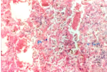
One hundred human foetuses having crown-rump length in the range of 7–36 cm and gestational age of 12–36 weeks were subjected to morphological and histological studies to understand and observe the developing medulla of foetal adrenal glands. It is observed that from week 12 to 14, medulla is not clearly demarcated and clusters of medullary cells are found in the foetal cortex. Towards week 38, we observe that the medulla is well organized and differentiated with phaechromocytes and few ganglion cells clearly visible. The observations are important for clinicians to help in diagnosis of neuroblastomas.
References
No records found.
About this article

Cite this article
Licence:
| Received | Accepted | Published |
|---|---|---|
| N/A | N/A | N/A |
DOI: http://dx.doi.org/10.21088/ija.2320.0022.7218.3
Keywords
Medulla; Foetal Adrenal Glands; Neuroblasts; Phaeochromocytes.Search for Similar Articles
Similar Articles
- Supernumerary Pulmonary Vein: A Rare Anatomical Variation Observed During Cadave...
- Elongated Styloid Process: Anatomical Variations and Their Clinical Relevance: A...
- Bridging the Gap Between Dissection and Surgery: The Role of Soft Embalming in M...
- A Cadaveric Study of Retro Hepatic Segment of Inferior Vena Cava and Ostia Venae...
- Estimation of Stature from Forearm Length in Medical Undergraduate Students of a...
Article Level Metrics
Last UpdatedSunday 20 September 2026, 21:53:10 (IST)
5038
Accesses
9
601
00
Citations
NA
NA
NA
Download citation
Article Keywords
Keyword Highlighting
Highlight selected keywords in the article text.
Timeline
| Received | N/A |
| Accepted | N/A |
| Published | N/A |