Full Text (PDF)
Indian Journal of Forensic Medicine and Pathology 16(1):p 73-76, January-March 2023. | DOI: https://doi.org/10.21088/ijfmp.0974.3383.16123.9
Case Report
Leiomyoma of Broad Ligament a Location with Rare Incidence: A Case Report
Bhavinee Pathak, Sabiha Maimoon, Kalpana A. Bothale
Author Information
Licence:
Attribution-Non-commercial 4.0 International (CC BY-NC 4.0)This license enables reusers to distribute, remix, adapt, and build upon the material in any medium or format for noncommercial purposes only, and only so long as attribution is given to the creator.
Indian Journal of Forensic Medicine and Pathology 16(1):p 73-76, January-March 2023. | DOI: https://doi.org/10.21088/ijfmp.0974.3383.16123.9
How Cite This Article:
Pathak B, Maimoon S, Bothale KA. Leiomyoma of broad ligament (a location with rare incidence): a case report. Indian J Forensic Med Pathol. 2023;16(1):73-76.Timeline
Received : October 17, 2022
Accepted : December 13, 2022
Published : March 30, 2023
Abstract
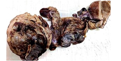
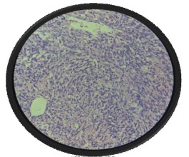
Leiomyoma are most common benign tumors of the uterus, mostly situated in the body of the uterus. Rarely, they arise from extrauterine sites with broad ligament leiomyoma being uncommon.1 A 49-year-old female, multipara presented in outpatient department with complaints of lower abdominal pain and abdominal distension. Abdominal examination revealed a firm mass arising from the pelvis corresponding to 18 week size. On per vaginal examination, huge mass was palpable occupying whole abdomen; uterus was not felt separately. CECT abdomen and pelvis revealed lesion arising from right adnexa and occupying pelvic region most likely suggestive of neoplastic etiology of ovary? USG was suggestive of an abdominopelvic neoplastic lesion arising from right adnexa. Total abdominal hysterectomy with bilateral salpingo-oophorectomy was done. The specimen was sent for histopathological examination. Histopathological examination revealed broad ligament leiomyoma. This case is being reported for its rare incidence, diagnostic dilemma with ovarian malignancy and surgical challenge.
References
- 1. Purwar N, Dwivedi S, Tripathi BN. A huge broad ligament fibroid with degenerative changes: a rare case report. Journal of Evolution of Medical and Dental Sciences. 2016 May 5;5(36):2171–3.
- 2. Guo L, Wang MZ, Wang LJ, Zang AH, Jia DM, Hou L, et al. Giant broad ligament leiomyoma with postoperative infection diagnosed with contrast-enhanced ultrasound: Case report and literature review. J Clin Ultrasound. 2022 Jan;50(1):138–47.
- 3. Podduturi V, Armstrong-Briley DR, Guileyardo JM. Sudden Death by Pulmonary Thromboembolism due to a Large Uterine Leiomyoma with a Parasitic Vein to the Mesentery. Case Rep Obstet Gynecol. 2014;2014:181265.
- 4. A Large Cystic Degenerating Broad ligament Leiomyoma Masquerading as Ovarian Malignancy! [Internet]. [cited 2022 Apr 7]. Available from: http://www.casereports. i n / a r t i c l e s / 5 / 2 / A - L a rg e - Cy s t i c - D e g e n e ra t i n g - B ro a d - l i g a m e n t - Le i o m y o m a - M a s q u e ra d i n g - a s - Ovarian-Malignancy.html
- 5. Fasih N, Prasad Shanbhogue AK, Macdonald DB, Fraser-Hill MA, Papadatos D, Kielar AZ, et al. Leiomyomas beyond the uterus: unusual locations, rare manifestations. Radiographics. 2008 Dec;28(7):1931–48.
- 6. Vitthalrao JS, Anita G. Broad Ligament Leiomyoma Mimicking As Ovarian Tumor. :3.
- 7. Parasitic and pedunculated leiomyomas: ultrasonographic features. - Yeh - 1999 - Journal of Ultrasound in Medicine - Wiley Online Library [Internet]. [cited 2022 Apr 6]. Available from: https://onlinelibrary. w i l e y . c o m / d o i / a b s / 1 0 . 7 8 6 3 / jum.1999.18.11.789
- 8. Dekel A, Rabinerson D, Dicker D, Ben-Rafael Z. Thrombosis of the pelvic veins associated with a large myomatous uterus. Obstet Gynecol. 1998 Oct;92(4 Pt 2):646–7.
- 9. Jacobs I, Bast RC. The CA 125 tumour-associated antigen: a review of the literature. Hum Reprod. 1989 Jan;4(1):1–12.
Data Sharing Statement
There are no additional data available. All raw data and code are available upon request.
Funding
This research received no funding.
Author Contributions
All authors contributed significantly to the work and approve its publication.
Ethics Declaration
This article does not involve any human or animal subjects, and therefore does not require ethics approval.
Acknowledgements
We would like to express our gratitude to the patients, their families, and all those who have contributed to this study.
Conflicts of Interest
No conflicts of interest in this work.
About this article

Cite this article
Pathak B, Maimoon S, Bothale KA. Leiomyoma of broad ligament (a location with rare incidence): a case report. Indian J Forensic Med Pathol. 2023;16(1):73-76.
Licence:
Attribution-Non-commercial 4.0 International (CC BY-NC 4.0)This license enables reusers to distribute, remix, adapt, and build upon the material in any medium or format for noncommercial purposes only, and only so long as attribution is given to the creator.
| Received | Accepted | Published |
|---|---|---|
| October 17, 2022 | December 13, 2022 | March 30, 2023 |
DOI: https://doi.org/10.21088/ijfmp.0974.3383.16123.9
Keywords
Broad ligamentLeiomyomaOvarian malignancycase report.LeiomyomaOvarian malignancycase report.Search for Similar Articles
Similar Articles
- Diatom Analysis in Thermophilic water bodies: A Study on Hot Water Springs of Ma...
- Morphometric Study of Supra-meatal Triangle and Mastoid process in the Adult dry...
- Histopathological Spectrum of Autopsy Cases: A One Year Retrospective Observatio...
- Forensic Differentiation of Manner of Death in Railway Fatalities: A FourCase Se...
- Understanding assault Patterns: A Cross-sectional study of Cases at Chri, Kelamb...
Article Level Metrics
Last UpdatedMonday 19 October 2026, 10:56:30 (IST)
16327
Accesses
44
3215
00
Citations
NA
NA
NA
Download citation
Article Keywords
Keyword Highlighting
Highlight selected keywords in the article text.
Timeline
| Received | October 17, 2022 |
| Accepted | December 13, 2022 |
| Published | March 30, 2023 |
licence
Attribution-Non-commercial 4.0 International (CC BY-NC 4.0)
This license enables reusers to distribute, remix, adapt, and build upon the material in any medium or format for noncommercial purposes only, and only so long as attribution is given to the creator.