Full Text (PDF)
Indian Journal of Forensic Medicine and Pathology 18(1):p 35-45, Jan -March 2025. | DOI: https://doi.org/10.21088/ijfmp.0974.3383.18125.5
Original Article
Investigating the Association between Palatal Rugae and Sagittal Malocclusion: A Clinical Study
Poorna Devadoss, Ramachandra Prabhakar, Umamaheshwari R., Saravanan R., Sagaya Mary Priya R. P., Sameera Y.
Author Information
Licence:
Attribution-Non-commercial 4.0 International (CC BY-NC 4.0)This license enables reusers to distribute, remix, adapt,
and build upon the material in any medium or format for noncommercial purposes
only, and only so long as attribution is given to the creator.
Indian Journal of Forensic Medicine and Pathology 18(1):p 35-45, Jan -March 2025. | DOI: https://doi.org/10.21088/ijfmp.0974.3383.18125.5
How Cite This Article:
Poorna Devadoss, Ramachandra Prabhakar, et al. Investigating the Association Between Palatal Rugae Patterns and Sagittal Malocclusion: A Clinical Study. Indian J Forensic Med Pathol. 2025;18(1):35-45.Timeline
Received : January 03, 2025
Accepted : March 06, 2025
Published : March 25, 2025
Abstract
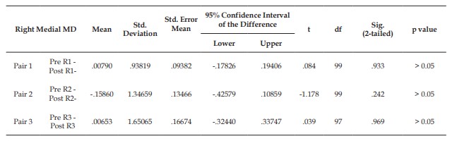
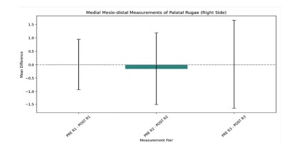
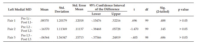
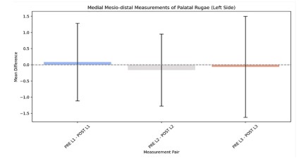
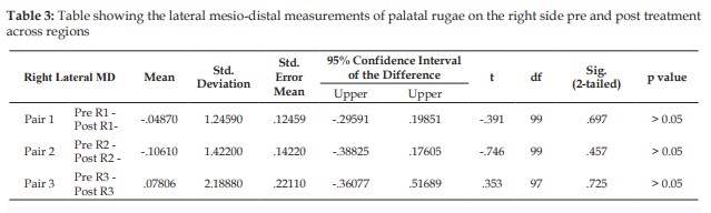
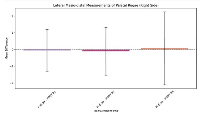
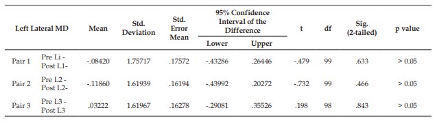
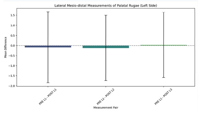
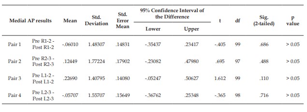
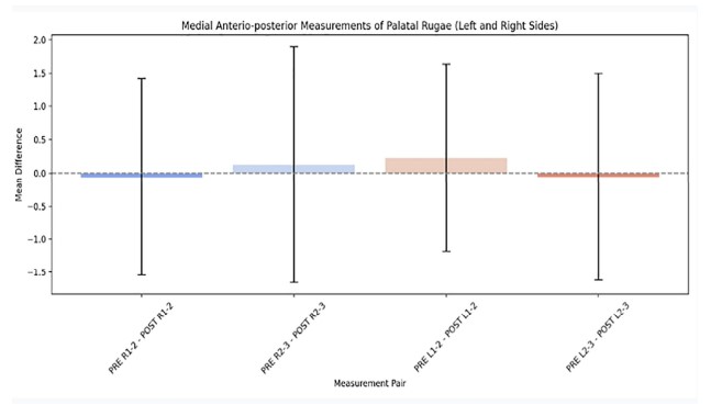
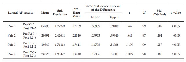
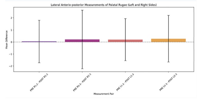
Introduction: Palatal rugae are unique anatomical ridges located on the anterior hard palate, remaining stable throughout an individual’s life. In forensic odontology and orthodontics, they serve as key landmarks. The stability of palatal rugae, coupled with their resistance to change, suggests potential diagnostic value, especially in sagittal malocclusion classified as Class I, II, or III by Angle’s system. This study investigates the association between palatal rugae patterns and sagittal malocclusion, exploring whether their morphological characteristics can aid in malocclusion diagnosis. Objectives: To examine whether palatal rugae patterns (length and width) are significantly associated with sagittal malocclusions (Class I, II, and III) and to assess their potential role as non-invasive diagnostic tools for orthodontic assessment. Methodology: This cross-sectional clinical study involved 300 participants, aged 18 to 28, divided equally into three groups based on untreated sagittal malocclusion: Class I, Class II, and Class III, according to Angle’s classification. Strict inclusion criteria ensured participants had no history of orthodontic treatment, systemic diseases, facial trauma, or habits affecting palatal anatomy. Pre-treatment maxillary dental models were made using high-quality alginate impressions and dental stone casts, focusing on primary palatal rugae for analysis. Key parameters measured were the mesio-distal (MD) length and anterio-posterior (AP) width of the rugae, with data collected using digital calipers. Statistical analysis was conducted with SPSS Ver 26.0, employing ANOVA to compare rugae dimensions between malocclusion groups, and Pearson Correlation Coefficient to assess the relationship between rugae patterns and malocclusion. A significance level of p<0.05 was set. Results: The paired sample t-test results revealed no statistically significant difference in palatal rugae dimensions before and after orthodontic treatment across the regions studied (right and left medial and lateral rugae). For mesiodistal (MD) measurements, the mean difference for right-side rugae ranged from -0.0487 to 0.07806, while left-side rugae differences ranged from -0.0842 to 0.0837, with p-values consistently greater than 0.05. Similarly, for anterio-posterior (AP) measurements, the mean difference ranged from -0.0601 to 0.2622 across both sides, with no significant p-values observed. These findings confirm that palatal rugae patterns remained stable post-treatment, indicating no measurable changes in their dimensions. Conclusion: Palatal rugae patterns are not significantly associated with sagittal malocclusion, and their dimensions remain stable following orthodontic interventions. They can serve as reliable anatomical landmarks in orthodontics and forensic identification, offering potential for non-invasive diagnostics in clinical settings.
References
- 1. 2. Shailaja AM, Romana IRU, Narayanappa G, Smitha T, Gowda NC, Vedavathi HK. Assessment of palatal rugae pattern and its significance in orthodontics and forensic odontology. J Oral Maxillofac Pathol. 2018;22(3):430-435.
- 2. Ziar N, Pakshir HR, Alamdarloo Y, et al. Characteristic changes of the palatal rugae following orthodontic treatment. Egypt J Forensic Sci. 2023;13(14).
- 3. Pazera C, Gkantidis N. Palatal rugae positional changes during orthodontic treatment of growing patients. Orthod Craniofac Res. 2021;24(3):351-359.
- 4. Mustafa AG, Allouh MZ, Alshehab RM. Morphological changes in palatal rugae patterns following orthodontic treatment. J Forensic Leg Med. 2015;31:19-22.
- 5. Lanteri V, Cossellu G, Farronato M, et al. Assessment of the Stability of the Palatal Rugae in a 3D-3D Superimposition Technique Following Slow Maxillary Expansion (SME). Sci Rep. 2020;10:2676.
- 6. Alshammari A, Farook FF, Alyahya L, et al. A Morphometric Analysis of Palatal Rugae Patterns in a Saudi Arabian Population. Cureus. 2022;14(12):e33058.
- 7. Abdel Aziz H, Sabet N. Palatal rugae area: a landmark for analysis of pre- and post-orthodontically treated adult Egyptian patients. East Mediterr Health J. 2001;7(1-2):60-66.
- 8. Bansode SC, Kulkarni MM. Importance of palatal rugae in individual identification. J Forensic Dent Sci. 2009;1(2):77.
- 9. Bailey LTJ, Esmailnejad A, Almeida MA. Stability of the palatal rugae as landmarks for analysis of dental casts in extraction and nonextraction cases. Angle Orthod. 1996;66(1):73-78.
- 10. Damstra J, Mistry D, Cruz C, Ren Y. Antero-posterior and transverse changes in the positions of palatal rugae after rapid maxillary expansion. Eur J Orthod. 2009;31(3):327-332.
- 11. Deepak V, Malgaonkar NI, Shah NK, et al. Palatal rugae patterns in orthodontically treated cases, are they a reliable forensic marker? J Int Oral Health. 2014;6(5):89-95.
- 12. Gujar A, Rani M. Three-dimensional assessment of the palatal contour changes in orthodontically treated cases: A scanned maxillary cast analysis. J Indian Orthod Soc. 2016;50(3):145-149.
- 13. Hoggan BR, Sadowsky C. The use of palatal rugae for the assessment of anteroposterior tooth movements. Am J Orthod Dentofacial Orthop. 2001;119(5):482-488.
- 14. Kapoor P, Miglani R. Transverse changes in lateral and medial aspects of palatal rugae after mid palatal expansion: a pilot study. J Forensic Dent Sci. 2015;7(1):8-13.
- 15. Sivaraj A. Significance of Palatal Rugae in Orthodontics. J Orofac Res. 2013;3(3):202-209.
- 16. Tey SN, Lin YM, Syed Mohamed AMF. Stability of palatal rugae after orthodontic/orthopaedic expansion: a scoping review. Aust Orthod J. 2023;39:158-170.
Data Sharing Statement
There are no additional data available.
Funding
This research received no funding.
Author Contributions
All authors contributed significantly to the work and approve its publication.
Ethics Declaration
his article does not involve any human or animal subjects, and therefore does not require ethics approval.
Acknowledgements
Information not provided.
Conflicts of Interest
The authors report no conflicts of interest in this work.
About this article
Cite this article
Poorna Devadoss, Ramachandra Prabhakar, et al. Investigating the Association Between Palatal Rugae Patterns and Sagittal Malocclusion: A Clinical Study. Indian J Forensic Med Pathol. 2025;18(1):35-45.
Licence:
Attribution-Non-commercial 4.0 International (CC BY-NC 4.0)This license enables reusers to distribute, remix, adapt,
and build upon the material in any medium or format for noncommercial purposes
only, and only so long as attribution is given to the creator.
| Received | Accepted | Published |
|---|---|---|
| January 03, 2025 | March 06, 2025 | March 25, 2025 |
DOI: https://doi.org/10.21088/ijfmp.0974.3383.18125.5
Keywords
Palatal rugaeSagittal malocclusionClass-I malocclusionClass-II malocclusionClass-III malocclusionOrthodonticsForensic odontologyPalatal rugoscopySearch for Similar Articles
Similar Articles
- Pesticide Contamination in Indian Agricultural and Residential Areas: A Compara...
- Forensic Age Estimation using CBCT-Derived Mandibular Morphometrics: A Comparat...
- A 2 Years Retrospective Study of the Spectrum of Poisoning in a Tertiary Care C...
- Knowledge and Attitude of MBBS Students Regarding Post Mortem Examination: A Cr...
- Sudden Death in a 7-year-old Child Due to Neoplasm: A Case Report
Article Level Metrics
Last UpdatedWednesday 08 July 2026, 06:33:19 (IST)
7722
Accesses
31
2137
00
Citations
NA
NA
NA
Download citation
Article Keywords
Keyword Highlighting
Highlight selected keywords in the article text.
Timeline
| Received | January 03, 2025 |
| Accepted | March 06, 2025 |
| Published | March 25, 2025 |
licence
Attribution-Non-commercial 4.0 International (CC BY-NC 4.0)
This license enables reusers to distribute, remix, adapt,
and build upon the material in any medium or format for noncommercial purposes
only, and only so long as attribution is given to the creator.