Full Text (PDF)
Indian Journal of Forensic Medicine and Pathology 15(4):p 255-262, October-December 2022. | DOI: https://doi.org/10.21088/ijfmp.0974.3383.15422.8
Original Article
Histopathological Spectrum of Appendicectomy Specimens with Emphasis on Unusual Findings
Shruti Sainath Andola, Sainath K Andola, Priyanka Patil, Rohit Patil
Author Information
Licence:
Attribution-Non-commercial 4.0 International (CC BY-NC 4.0)This license enables reusers to distribute, remix, adapt, and build upon the material in any medium or format for noncommercial purposes only, and only so long as attribution is given to the creator
Indian Journal of Forensic Medicine and Pathology 15(4):p 255-262, October-December 2022. | DOI: https://doi.org/10.21088/ijfmp.0974.3383.15422.8
How Cite This Article:
Andola SK, Patil P, Patil R, et al. Histopathological spectrum of appendicectomy specimens with emphasis on unusual findings. Indian J Forensic Med Pathol. 2022;15(4):255-262.Timeline
Received : June 10, 2022
Accepted : October 11, 2022
Published : December 30, 2022
Abstract
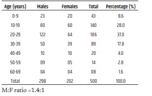
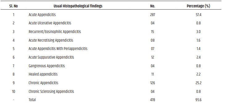
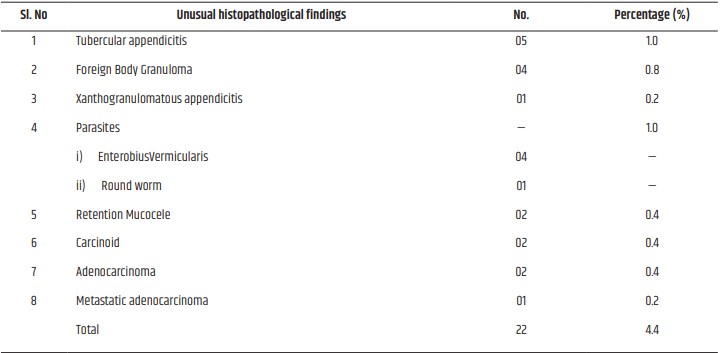
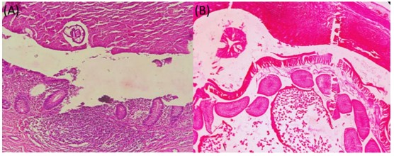
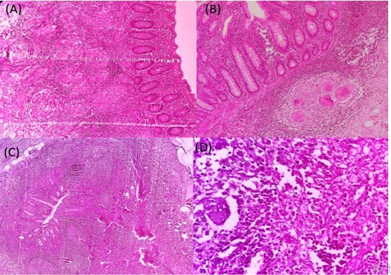
Introduction: The present study was carried out to assess the value of routine histopathological examination of appendectomy specimens An attempt is made to review the histopathological spectrum of appendectomy over a period of 2 years 3 months. Aims and Objectives: To study the spectrum of histopathological lesions in appendectomy specimens and special emphasis is made to highlight the unusual lesions observed in surgically resected appendicectomies and appendix in medicolegal autopsies. Material and Methods: The study was carried out on appendectomy specimens received in the department of pathology Mahadevappa Rampure Medical College during a period of 2 years 3 months from Jan 2019 to March 2021. Clinical data collected from records of corresponding appendectomy specimens were processed. Sections studied for various histopathological patterns with an emphasis on unusual findings. Results: A total of 500 specimens were analysed among which 298 were males and 202 were females with M:F 1.4:1. The histopathological examination showed Acute appendicitis (57.4%), Acute ulcerative appendicitis (0.8%), Recurrent/Eosinophilic appendicitis (03%), Acute necrotising appendicitis (1.6%), Acute appendicitis with periappendicitis (1.4%), Acute suppurative appendicitis (2.4%), Gangrenous appendicitis (0.8%), Healed appendicitis (2.2%), Chronic appendicitis (25.2%) and Chronic sclerosing appendicitis (1.0%). The unusual fingings were observed in 22 cases (4.4%) which include Tubercular appendicitis (1.0%), Foreign body granuloma (0.8%), Xanthogranulomatous appendicitis (0.2%), Enterobiusvermicularis (0.8%), Round worm (0.2%), Retention mucocele (0.4%), Carcinoid tumor (0.4%), Mucinous adenocarcinoma (0.4%) and Metastatic adenocarcinoma (0.2%). Conclusion: Though the present study revealed usual findings of appendicitis and variants in most of the cases, the observation of unusual findings like granulomas, parasites and neoplasms warrant a careful study of all appendectomy specimens received in pathology department with emphasis on relevant clinical and laboratory findings thus modulating the management and follow up
References
- 1. Marudanayagam R, Williams GT, Rees BI; Review of the pathological results of 2660 appendectomy specimens Gastro., 2006;41(8):745-749.
- 2. Oguntola AS, Adeoti ML, Oyemolade TA. Appendicitis: Trends in incidence, age, sex and seasonal variations in South-western Nigeria. Ann Afr Med 2010;9:213-217.
- 3. Turner JR. The Gastrointestinal tract, In: Kumar, Abbas, Fausto(eds). Robins and Cotran Pathologic basis of disease.8th edn. Saunders: Philadelphia;2010. pp870-871.
- 4. Fergusson JAE, Hitos K, Simpson E. Utility of white cell count and ultrasound in the diagnosis of acute appendicitis. ANZ J Surg 2002;72:781- 785.
- 5. O’Connell PR. The vermiform appendix. In: Russell RC, Williams NS, Bulstrode CJ. editors. Bailey and Love’s Short Practice of Surgery. 26th ed. London: Arnold Hodder; 2010.p.1203-1218.
- 6. Duzgun AP, Moran M, Uzun S, Ozmen MM, Ozer VM, Seckin S et al. Unusual findings in appendectomy specimens: Evaluation of 2458 cases and review of the literature. Indian J Surg 2004;66:221-226.
- 7. Zulfikar I, Khanzada TW, Sushel C, Samad A; Review of the pathologic diagnoses of appendectomy specimens. Annals of King Edward Medical University, 2009;15(4):168-170.
- 8. Blair NP, Bugis SP, Turner LJ, Macleod MM; Review of pathological diagnosis of 2216 appendectomy specimens. Am J Surg.,1993;165(5):618-620.
- 9. Edino ST, Mohammed AZ, Ochicha O, Anumah M; Appendicitis in Kano, Nigeria: A 5year review of pattern, morbidity and mortality. Annals of African Medicine, 2004;3(1):38-41.
- 10. Majid S, Imran AA Khan SA; Morphological variations in appendectomy specimens. Pak J Pathol., 2005;16(2):58-60.
- 11. Rabindranath D, Khan AA, Ansari H, Senthil P. Unusual incidental findings of routine histopathological examination of appendectomy specimens- a 2-year retrospective analysis with review of the literature. Int J of Allied Med Sci and Clin Res 2016; 4(1):90-98.
- 12. Abdulkarim Hasan1*, Khalid Mohamed Nafie2, Osama Sharafeldin Abbadi; The Utility of Routine Histopathological Examination of the Appendectomy Specimens; Annals of Pathology and Laboratory Medicine, Vol. 7, Issue 7, July, 2020.
- 13. Aneel Myageri1, Aditya Divakar Agnihotri2, Lokesh Durjan Singh Chauhan; Clinicopathologic Study of Appendix Specimens-A Two Year Retrospective Study at a Tertiary Care Center; National Journal of Laboratory Medicine. 2019 Apr, Vol-8(2): PO05- PO10.
- 14. Aravindan K. P., Deepthy Vijayaraghavan, Marie Therese Manipadam: Acute eosinophilic appendicitis and the significance of eosinophil-Edema lesion: indian journal of pathology an dmicrobiology-53(2), april-june 2010.
- 15. KP Aravindan, Deepthy Vijayaraghavan, Marie Therese Manipadam. Significance of eosinophil-edema lesion. Indian J Pathol Microbiol 2010;53:258-261.
- 16. Aravindan KP. Eosinophils in acute appendicitis: Possible significance. Indian J PatholMicrobiol 1997;40:491-498.
- 17. Rai SP, Shukla A, Kashyap M, Dahiya RK. Isolated tuberculosis of the appendix. Indian J Tuberc 2004;51:239-240.
- 18. Wani I, Maqbool M, Amin A, Shah F, Keema A, Singh J, et al. Appendicealascariasis in children. Ann Saudi Med 2010; 30:63-66.
- 19. In’t Hof KH, Van Der Wal HC, Kazemier G., Lange JF; Carcinoid tumor of the appendix. Analyses of 1485 consecutive emergency appendicectomies J GastrointestSurg 2008;12(8):1436- 1438.
- 20. Matthyssens LE, Ziol M, Barrat C, Champault GG: Routine Surgical Pathology in General Surgery.Br J Surg 2006;93:362-368.
- 21. Sieren LM, Collins JN, Weireter LJ, Britt RC, Reed SF, Novosel TJ, Britt LD. The incidence of benign and malignant neoplasia presenting as acute appendicitis. Am Surg 2010;76:808-811.
- 22. Cortina R, McCormick J, Kolm P, Perry RR. Management and prognosis of adenocarcinoma of the appendix.Dis Colon Rectum 1995;38:848.
- 23. Connor SJ, Hanna GB, Frizelle FA. Appendicealtumors: retrospective clinicopathologic analysis of appendicealtumors from 7,970 appendectomies. Dis Colon Rectum. 1998;41:75–80.
- 24. Mallikarjun Pattanashetti1, MM Priyadarshini2, BN Gayathri3; Histopathological Study of Appendicectomy Specimens; National Journal of Laboratory Medicine. 2021 Jan, Vol-10(1): PO30-PO33.
Data Sharing Statement
There are no additional data available. All raw data and code are available upon request.
Funding
This research received no funding.
Author Contributions
All authors contributed significantly to the work and approve its publication.
Ethics Declaration
This article does not involve any human or animal subjects, and therefore does not require ethics approval.
Acknowledgements
We would like to express our gratitude to the patients, their families, and all those who have contributed to this study.
Conflicts of Interest
No conflicts of interest in this work.
About this article
Cite this article
Andola SK, Patil P, Patil R, et al. Histopathological spectrum of appendicectomy specimens with emphasis on unusual findings. Indian J Forensic Med Pathol. 2022;15(4):255-262.
Licence:
Attribution-Non-commercial 4.0 International (CC BY-NC 4.0)This license enables reusers to distribute, remix, adapt, and build upon the material in any medium or format for noncommercial purposes only, and only so long as attribution is given to the creator
| Received | Accepted | Published |
|---|---|---|
| June 10, 2022 | October 11, 2022 | December 30, 2022 |
DOI: https://doi.org/10.21088/ijfmp.0974.3383.15422.8
Keywords
AppendectomyUnusual lesionsHistopathologyAppendicitis.Unusual lesionsHistopathologyAppendicitis.Search for Similar Articles
Similar Articles
- Sudden Death in a 7-year-old Child Due to Neoplasm: A Case Report
- Bites of Burden: Snake Bite Envenoming in India, Regional Venom Differences, Tr...
- Forensic View of Suicide in Healthcare Workers
- Tracing Identity from Teeth to Technology: Artificial Intelligence in Forensic...
- Chemical Constituents in Latent Fingermarks as Indicators of Lifestyle using Gas...
Article Level Metrics
Last UpdatedSunday 21 June 2026, 19:39:17 (IST)
7238
Accesses
8
2068
00
Citations
NA
NA
NA
Download citation
Article Keywords
Keyword Highlighting
Highlight selected keywords in the article text.
Timeline
| Received | June 10, 2022 |
| Accepted | October 11, 2022 |
| Published | December 30, 2022 |
licence
Attribution-Non-commercial 4.0 International (CC BY-NC 4.0)
This license enables reusers to distribute, remix, adapt, and build upon the material in any medium or format for noncommercial purposes only, and only so long as attribution is given to the creator