Full Text (PDF)
Indian Journal of Pathology: Research and Practice 11(3):p 91-94, July-September 2022. | DOI: https://doi.org/10.21088/ijprp.2278.148X.11322.9
Original Article
Fine Needle Aspiration Cytology of Salivary Gland Lesions with Application of Milan System of Reporting
Lakshmi Kanth Dhandapani, Nandini S, Smita S K, Deepa S Masur, S S Hiremath
Author Information
Licence:
Attribution-Non-commercial 4.0 International (CC BY-NC 4.0)This license enables reusers to distribute, remix, adapt, and build upon the material in any medium or format for noncommercial purposes only, and only so long as attribution is given to the creator.
Indian Journal of Pathology: Research and Practice 11(3):p 91-94, July-September 2022. | DOI: https://doi.org/10.21088/ijprp.2278.148X.11322.9
How Cite This Article:
Lakshmi Kanth Dhandapani, Nandini S, Smita S K et al. / Fine Needle Aspiration Cytology of Salivary Gland Lesions with Application of Milan System of Reporting./Indian Journal of Pathology: Research and Practice Volume 11 Number 3, July–Sept 2022. 91–94.Timeline
Received : February 08, 2022
Accepted : March 05, 2022
Published : September 25, 2022
Abstract
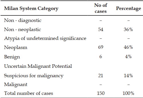
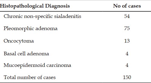
Introduction: FNAC of salivary is simple, easy, cost-effective investigation that can be used to differentiate benign and malignant lesions. Milan system was introduced to improve the communication between cytopathologists and physicians. Objectives: To diagnose the salivary gland swelling using fine needle aspirations by Milan system of reporting and correlate with histopathology. Materials and methods: Total number of 150 cases of salivary gland lesions were studied from September 2020 to August 2021 in SNMC and HSK hospital. Smears were then stained with H&E, Giemsa and PAP stains. Results: A total 150 patients that underwent FNAC, reported using Milan system of reporting and followed up with histopathological correlation. Results revealed that FNAC was effective in differentiating benign and malignant conditions by using Milan system of reporting. Conclusion: FNAC is a safe, reliable and yet economically effective technique in diagnosing salivary gland lesions. Milan system of reporting is essential to improve the communication between cytopathologists and physicians and gives universal approach in diagnosis and management of salivary gland lesions. So FNAC with Milan system can be used as effective pre-operative investigation in patients with salivary gland lesions.
References
- 1. Song SJ, Shafique K, Wong LQ, LiVolsi VA, Montone KT, Baloch Z. The utility of the Milan System as a risk stratification tool for salivary gland fine needle aspiration cytology specimens. Cytopathology.2019.
- 2. Pujani M, Chauhan V, Agarwal C, Raychaudhuri S, Singh K. A critical appraisal ofthe Milan system for reporting salivary gland cytology (MSRSGC) with histological correlation over a 3-year period: Indian scenario. DiagnCytopathol.2019.
- 3. Jalaly JB, Farahani SJ, Baloch ZW. The Milan system for reporting salivary gland cytopathology: A comprehensive review of the literature. Diagnostic Cytopathology. 2020:1-10.
- 4. Faquin WC, Rossi ED et al. The Milan System for Reporting Salivary Gland Cytopathology. Springer International Publishing AG,2018;p1-11,157-169.
- 5. Pusztaszeri M, Rossi ED, Baloch ZW, Faquin WC. Salivary Gland Fine Needle Aspiration and Introduction of the Milan Reporting System. AdvAnatPathol. 2019 Mar;26(2):84-92
- 6. Wei S, Layfield LJ, LiVolsi VA, Montone KT, Baloch ZW. Reporting of fine needle aspiration (FNA) specimens of salivary gland lesions: A comprehensive review. DiagnCytopathol. 2017Sep;45(9):820-827.
- 7. Mukundapai M, Sharma N, Patil A, Gopal C. Fineneedle Aspiration Cytology of Salivary Gland Lesions: A revised classification based on “Milan System” – 4 years experience of Tertiary care cancer center of South India. J cytol.2020;37(1):12-17.
- 8. Mullen D. Gibbons D. A retrospective comparison of salivary gland fine needleaspiration reporting with the Milan system for reporting salivary gland cytology. Cytopathology. 2020;31:208-214.
- 9. Viswanathan K, Sung S, Scognamiglio T, Yang GC, Siddiqui MT, Rao RA. The role of the Milan system for reporting salivary gland cytopathology: a 5-year institutional experience. CancerCytopath ol.2018;126(8):541-551.
- 10. Wu HH, Alruwaii F, Zeng B-R, Cramer HM, Lai C-R, Hang J-F. Application of the Milan system for reporting salivary gland cytopathology: a retrospective 12-year bi-institutional study. Am J ClinPathol.2019;151(6):613-621.
Data Sharing Statement
There are no additional data available. All raw data and code are available upon request.
Funding
This research received no funding.
Author Contributions
All authors contributed significantly to the work and approve its publication.
Ethics Declaration
This article does not involve any human or animal subjects, and therefore does not require ethics approval.
Acknowledgements
We would like to express our gratitude to the patients, their families, and all those who have contributed to this study.
Conflicts of Interest
No conflicts of interest in this work.
About this article

Cite this article
Lakshmi Kanth Dhandapani, Nandini S, Smita S K et al. / Fine Needle Aspiration Cytology of Salivary Gland Lesions with Application of Milan System of Reporting./Indian Journal of Pathology: Research and Practice Volume 11 Number 3, July–Sept 2022. 91–94.
Licence:
Attribution-Non-commercial 4.0 International (CC BY-NC 4.0)This license enables reusers to distribute, remix, adapt, and build upon the material in any medium or format for noncommercial purposes only, and only so long as attribution is given to the creator.
| Received | Accepted | Published |
|---|---|---|
| February 08, 2022 | March 05, 2022 | September 25, 2022 |
DOI: https://doi.org/10.21088/ijprp.2278.148X.11322.9
Keywords
FNACMilan systemCytopathologistSearch for Similar Articles
Similar Articles
- Clinico-Pathological and Immunohistochemical Study of Primary CNS Tumors in a Te...
- The Role of SGLT2 Inhibitors in Cardiovascular Protection Beyond Diabetes: A Co...
- Epidermal Inclusion Cyst Presenting as 4th Ventricle Space Occupying Lesion: A R...
- Silent Retroperitoneal Seminoma Arising from an Occult Undescended Testis: Diagn...
- Scapular Metastasis of Oral Squamous Cell Carcinoma: A Case Report of an Unusual...
Article Level Metrics
Last UpdatedSaturday 17 October 2026, 10:30:48 (IST)
4177
Accesses
7
671
00
Citations
NA
NA
NA
Download citation
Article Keywords
Keyword Highlighting
Highlight selected keywords in the article text.
Timeline
| Received | February 08, 2022 |
| Accepted | March 05, 2022 |
| Published | September 25, 2022 |
licence
Attribution-Non-commercial 4.0 International (CC BY-NC 4.0)
This license enables reusers to distribute, remix, adapt, and build upon the material in any medium or format for noncommercial purposes only, and only so long as attribution is given to the creator.