Full Text (PDF)
Indian Journal of Pathology: Research and Practice 11(3):p 105-109, July-September 2022. | DOI: https://doi.org/10.21088/ijprp.2278.148X.11322.11
Original Article
Diagnostic Pitfalls in Salivary Gland Cytopathology with Emphasis on Milan System of Reporting
Author Information
Licence:
Attribution-Non-commercial 4.0 International (CC BY-NC 4.0)This license enables reusers to distribute, remix, adapt, and build upon the material in any medium or format for noncommercial purposes only, and only so long as attribution is given to the creator.
Indian Journal of Pathology: Research and Practice 11(3):p 105-109, July-September 2022. | DOI: https://doi.org/10.21088/ijprp.2278.148X.11322.11
How Cite This Article:
Rini Bishnoi/Diagnostic Pitfalls in Salivary Gland Cytopathology with Emphasis on Milan System of Reporting/Indian Journal of Pathology: Research and Practice 2022;11(3):105–109.Timeline
Received : January 08, 2022
Accepted : February 25, 2022
Published : September 25, 2022
Abstract
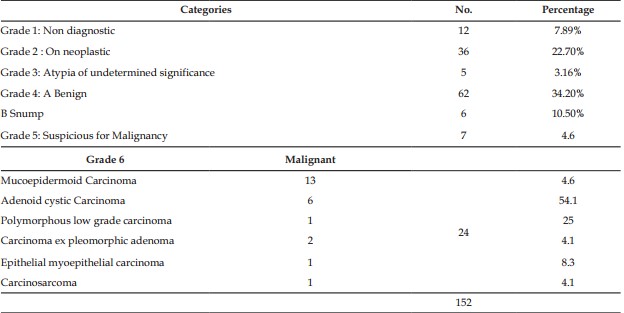
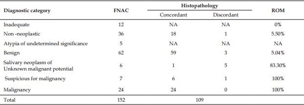
Salivary gland lesions are relatively less common but challenging because of their marked variability in cytomorphology, clinical features and biologic behavior. Fine needle aspiration cytology (FNAC) is helpful in evaluating suspicious salivary glands lesions due to its their superficial location, easy accessibility, low cost, minimum morbidity, rapid turnaround time, high specificity and sensitivity. Fine needle aspiration cytology in salivary gland lesions is intriguing for the cytopathologists due to their diverse morphology and overlapping cytological features which lead to the dilemma in diagnosis. Materials and Methods: Present study was a retrospective analytical study in a tertiary care centre over a duration of 3 years. All the salivary gland FNAC cases were retrieved and divided into various categories as per Milan system for reporting salivary gland cytopathology. Histopathological correlation was done wherever possible. Results: A total of 152 were included in the study. The percentage of cases in each category was: nondiagnostic 4.5%, nonneoplastic 51.9%, atypical lesions 0.76%, neoplastic category benign neoplasm 21.37%, salivary lesion of uncertain malignant potential 1.52%, suspicious category 2.29%, and malignant category 17.5%. The risk of malignancy for indivdual category was 6.25% (nonneoplastic), 100% (atypical), 3.3% (neoplastic), 0% (benign), 25% (salivary neoplasm of uncertain neoplastic potential), 100% (suspicious for malignancy), and 100% (malignant) categories. Sensitivity, specificity, positive predictive value, and negative predictive value of FNAC with application of Milan system was 89.4%, 100%, 100%, and 95.74%, respectively. Conclusion: Salivary gland FNAC when classified toa particular category by Milan system helps to overcome the pitfalls due to congruent features of various diagnostic categories yet provide risk stratification and stochastic information for the doubtful cases.
References
- 1. Barnes L, Eveson JW , Reichart P. , Sidransky D : Pathology and genetics of head and neck tumors in World Health Organization Classification of Tumors, P. Kleihues and L. H. Sobin, Eds., IARC Press, Lyon, France 2005 ; 210
- 2. A. A. Choudhury, T. Sultana, B. H. Siddique, and A. S. A. Amin, “Diagnosis of parotid gland mass by the fine needle aspiration cytology (FNAC) and its histopathological correlation—2 years study in BSMMU, Dhaka,” Bangabandhu Sheikh Mujib Medical University Journal, vol. 4, no. 2, pp. 65–69, 2011.
- 3. Frable MAS and FrableWJ :Fine-needle aspiration biopsy of salivary glands Laryngoscope,1991; 101: 245–249.
- 4. Mairembam P, Jay A, Beale T, et al. Salivary gland FNA cytology: role as a triage tool and an approach to pitfalls in cytomorphology. Cytopathology. 2016; 27: 91- 96.
- 5. Rajwanshi A, Gupta K, Gupta N, et al. Fine-needle aspiration cytology of salivary glands: diagnostic pitfalls—revisited. DiagnCytopathol. 2006; 34: 580-584.
- 6. Kim BY, Hyeon J, Ryu G, et al. Diagnostic accuracy of fine needle aspiration cytology for high-grade salivary gland tumors. Ann SurgOncol. 2013; 20: 2380- 2387.
- 7. Pastore A, Borin M, Malagutti N, et al. Preoperative assessment of salivary gland neoplasms with fine needle aspiration cytology and echography: a retrospective analysis of 357 cases. Int J ImmunopatholPharmacol. 2013; 26: 965- 971.
- 8. Postema RJ, van Velthuysen ML, van den Brekel MW, Balm AJ, Peterse JL. Accuracy of fine-needle aspiration cytology of salivary gland lesions in the Netherlands Cancer Institute. Head Neck. 2004; 26: 418- 424.
- 9. Stewart CJ, MacKenzie K, McGarry GW, Mowat A. Fine-needle aspiration cytology of salivary gland: a review of 341 cases. DiagnCytopathol. 2000; 22: 139-146.
- 10. Rossi ED, Wong LQ, Bizzarro T, Petrone G, Mule A, Fadda G, et al. The impact of FNAC in the management of salivary gland lesions: Institutional experiences leading to a risk based classification scheme. Cancer Cytopathol 2016;124:388 96.
- 11. Rohilla M, Singh P, Rajwanshi A, Gupta N, Srinivasan R, Dey P, et al. Three year cytohistological correlation of salivary gland FNA cytology at a tertiary center with the application of the Milan system for risk stratification. Cancer Cytopathol 2017;125:767 75
- 12. Lewis JE, Olsen KD, Sebo TJ. Carcinoma ex pleomorphic adenoma: pathological analysis of 73 cases. Hum Pathol. 2001;32:596–604
- 13. Orell S, Sterrett G. Orell, Orell and Sterrett's Fine Needle Aspiration Cytology. London: Elsevier Health Sciences UK; 2011.
- 14. Griffith CC, Pai RK, Schneider F, Duvvuri U, Ferris RL, Johnson JT, et al. Salivary gland tumor fine needle aspiration cytology: A proposal for a risk stratification classification. Am J ClinPathol 2015;143:839 53.
- 15. Amita K, Rakshitha HB, Singh A, Shankar SV. Evaluation of accuracy of milan system for reporting salivary gland cytology: Review of morphology and diagnostic challenges in each category. J Cytol 2020;37:18-25.
- 16. Kwon MY, Gu M. True malignant mixed tumor (carcinosarcoma) of parotid gland with unusual mesenchymal component. A case report and review of the literature. Arch Pathol Lab Med 2001; 125:812–5.
- 17. Sironi M, Isimbaldi G, Claren R, et al. Carcinosarcoma of the parotid gland: cytological, clinicopathological and immunohistochemical study of a case. Pathol Res Practice2000;196:511–517.
- 18. Nagel H, Laskawi R, Büter JJ, Schröder M, Chilla R, Droese M. Cytologic diagnosis of acinic cell carcinoma of salivary glands. DiagnCytopathol 1997;16:402 12
Data Sharing Statement
There are no additional data available. All raw data and code are available upon request.
Funding
This research received no funding.
Author Contributions
All authors contributed significantly to the work and approve its publication.
Ethics Declaration
This article does not involve any human or animal subjects, and therefore does not require ethics approval.
Acknowledgements
We would like to express our gratitude to the patients, their families, and all those who have contributed to this study.
Conflicts of Interest
No conflicts of interest in this work.
About this article

Cite this article
Rini Bishnoi/Diagnostic Pitfalls in Salivary Gland Cytopathology with Emphasis on Milan System of Reporting/Indian Journal of Pathology: Research and Practice 2022;11(3):105–109.
Licence:
Attribution-Non-commercial 4.0 International (CC BY-NC 4.0)This license enables reusers to distribute, remix, adapt, and build upon the material in any medium or format for noncommercial purposes only, and only so long as attribution is given to the creator.
| Received | Accepted | Published |
|---|---|---|
| January 08, 2022 | February 25, 2022 | September 25, 2022 |
DOI: https://doi.org/10.21088/ijprp.2278.148X.11322.11
Keywords
CytomorphologyDiagnostic accuracyFine needle aspiration cytologyRisk of malignancySalivary glandSearch for Similar Articles
Similar Articles
- Clinico-Pathological and Immunohistochemical Study of Primary CNS Tumors in a Te...
- The Role of SGLT2 Inhibitors in Cardiovascular Protection Beyond Diabetes: A Co...
- Epidermal Inclusion Cyst Presenting as 4th Ventricle Space Occupying Lesion: A R...
- Silent Retroperitoneal Seminoma Arising from an Occult Undescended Testis: Diagn...
- Scapular Metastasis of Oral Squamous Cell Carcinoma: A Case Report of an Unusual...
Article Level Metrics
Last UpdatedSaturday 17 October 2026, 10:30:07 (IST)
4177
Accesses
17
671
00
Citations
NA
NA
NA
Download citation
Article Keywords
Keyword Highlighting
Highlight selected keywords in the article text.
Timeline
| Received | January 08, 2022 |
| Accepted | February 25, 2022 |
| Published | September 25, 2022 |
licence
Attribution-Non-commercial 4.0 International (CC BY-NC 4.0)
This license enables reusers to distribute, remix, adapt, and build upon the material in any medium or format for noncommercial purposes only, and only so long as attribution is given to the creator.