Full Text (PDF)
Indian Journal of Anatomy 9(4):p 255-259, October-December 2020. | DOI: http://dx.doi.org/10.21088/ija.2320.0022.9420.4
Original Article
Clinical Anatomy of The Superficial Peroneal
Narendra A Patil. , Narendra A Patil1 , Mohammed Khaleel Ahmed2
Author Information
Licence:
Indian Journal of Anatomy 9(4):p 255-259, October-December 2020. | DOI: http://dx.doi.org/10.21088/ija.2320.0022.9420.4
How Cite This Article:
Narendra A Patil, Mohammed Khaleel Ahmed. Clinical Anatomy of The Superficial Peroneal. Indian J Anat. 2020;9(4):255–259.
Timeline
Received : N/A
Accepted : N/A
Published : N/A
Abstract
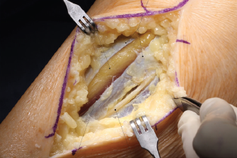
Background: Superficial perineal nerve (SPN): It is the nerve of lateral compartment of leg. It is one of the two terminal branches of the common perineal nerve given at the neck of the fibula, it arises in the substance of peroneus longus on the lateral side of the neck of fibula. Methods: A dissection was done in ten lower extremities (five from femaleand five from male) that had been well-maintained in a formalin arrangement. The subcutaneous layer was cautiously dissected in every preparation so that distortion of neural structures and their connections would be maintained a strategic distance from quite far. When the nerve structures had been recognized, the accompanying estimations were done: a) distance (in millimeters) from the upper end of the fibular head to the lower prominence of the lateral malleolus; b) number of branches from SPN when it arises through the superficial sural belt; c) distance (in millimeters) from the SPN rise highlight to the lateral malleolus; d) distance (in millimeters) from the SPN division to the lateral malleolus. Results: There were two primary discoveries from these dissections. To begin with, we noticed varieties in the quantity of branches passing through superficialanterior fascia of the leg. A few cases indicated a solitary trunk rise, later partitioning into two branches, while others demonstrated two diverse recently separated cutaneous branches. Second, in both anatomical sorts, a checked variety was seen in the relative distance from the lateral malleolus, emerging at a low, medium or elevated level. Regarding the compartment from which the SPN pierces the superficial fascia, the contrast between single-trunk and two branches ought to be considered. Conclusion: The information got appear to be adequately dependable for normal clinical practice, however it must be considered that successive and significant varieties may happen.
References
No records found.
About this article

Cite this article
Narendra A Patil, Mohammed Khaleel Ahmed. Clinical Anatomy of The Superficial Peroneal. Indian J Anat. 2020;9(4):255–259.
Licence:
| Received | Accepted | Published |
|---|---|---|
| N/A | N/A | N/A |
DOI: http://dx.doi.org/10.21088/ija.2320.0022.9420.4
Keywords
Surgical anatomy; Superficial peroneal nerve; Anatomical variations.Search for Similar Articles
Similar Articles
- Supernumerary Pulmonary Vein: A Rare Anatomical Variation Observed During Cadave...
- Elongated Styloid Process: Anatomical Variations and Their Clinical Relevance: A...
- Bridging the Gap Between Dissection and Surgery: The Role of Soft Embalming in M...
- A Cadaveric Study of Retro Hepatic Segment of Inferior Vena Cava and Ostia Venae...
- Estimation of Stature from Forearm Length in Medical Undergraduate Students of a...
Article Level Metrics
Last UpdatedTuesday 22 September 2026, 04:27:22 (IST)
5059
Accesses
6
655
00
Citations
NA
NA
NA
Download citation
Article Keywords
Keyword Highlighting
Highlight selected keywords in the article text.
Timeline
| Received | N/A |
| Accepted | N/A |
| Published | N/A |