Full Text (PDF)
Journal of Hospital Administration 6(1):p 27-31, January-June 2022. | DOI: NA
Case Report
Cardiac Complication of Extrapulmonary Tuberculosis
Abhishek Yadav, J N Pandit, Karthi null, Gokul null, Sudheer Arava
Author Information
Licence:
Attribution-Non-commercial 4.0 International (CC BY-NC 4.0)This license enables reusers to distribute, remix, adapt, and build upon the material in any medium or format for noncommercial purposes only, and only so long as attribution is given to the creator.
Journal of Hospital Administration 6(1):p 27-31, January-June 2022. | DOI: NA
How Cite This Article:
J. N. Pandit, Karthi, Gokul, et al./Cardiac Complication of Extrapulmonary Tuberculosis/RFP Journal of Hospital Administration. 2022;6(1):27-31.Timeline
Received : February 11, 2022
Accepted : May 24, 2022
Published : June 30, 2022
Abstract
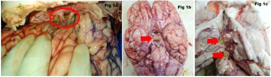
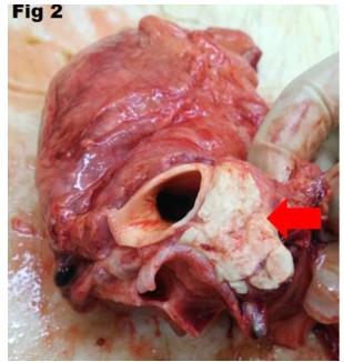
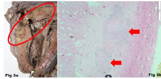
Among communicable diseases, tuberculosis (TB) is the major cluster causing mortality in young patients. WHO reported a global estimation of 10 million people fell ill with tuberculosis (1.5 million fatalities in 2020), and India alone contributed about26%.The incidence of Extra pulmonary Tuberculosis (ETB) is about 15% of all TB cases in India. Central nervous system (CNS) tuberculosis accounted for approximately 1% of total tuberculosis. TB is a rapidly progressive and fatal disease if left untreated. The progression of the disease and the occurrence of complications vary from patient to patient. Here the authors present a case of a young adult who presented with acute onset of fever and headache and she expired within a span of 31 days after the symptoms began. The novelty in the case is that an undiagnosed rare cardiac complication of extra pulmonary tuberculosis was diagnosed at autopsy. The investigations prior to her death didn’t indicate TB, but the treating physician had suspected ETB. The autopsy examination and histopathological examination supported the physician’s diagnosis. The authors had deliberated the pathogenesis of the cardiac complication of extra pulmonary tuberculosis contributing to the death of the deceased.
References
- 1. World Health Organization. Tuberculosis; Key facts. Internet. [accessed on 02.06.2022]. Available from. https://www.who.int/news-room/factsheets/detail/tuberculosis.
- 2. Ekaterina K, Kurt G N, Truls E B Johansen. Epidemiology of Extra pulmonary Tuberculosis. Chapter1.January2019.
- 3. Ingole R, Garg RK, Malhotra HS, Jain A, Kumar N, Rizvi I, Garg R. Spectrum of central nervous system tuberculosis: Anexperience from a large tertiary care institution of India. Indian JTuberc.2019;66:49–57.
- 4. Erbes R,Oettel K, Raffenberg M. Characteristics and outcome of patients with active pulmonary tuberculosis requiring intensive care. EurRespir J. 2006;27:1223–1228.
- 5. Lin CH, Lin CJ, KuoYW, Wang JY, Hsu CL, Chen JM, Cheng WC, Lee LN.Tuberculosis mortality: patient characteristics and causes. BMC Infect Dis. 2014;14:5
- 6. Abdelmalek R,Kanoun F, Kilani B, Tiouiri H, Zouiten F, Ghoubantini A, Chaanane T B. Tuberculous meningitis in adults: MRI contribution to the diagnosis in 29 patients. Int J Infect Dis. 2006;10:372-377
- 7. Kent SJ, Crowe SM, Yung A, Ron Lucas CM, Mijch A. Tuberculous meningitis: a 30-year review. Clin Infect Dis 1993;17:987-94.
- 8. Ortbals DW, Avioli LV. Tuberculous pericarditis. Arch Intern Med. 1979;139:231–234.
- 9. Mayosi BM, BurgessLJ, DoubellAF. Tuberculous pericarditis. Circulation. 2005;112:3608-3616.
- 10. Strang G, Latouf S, Commerford P, Roditi D, Traill D G, Barlow D, Forder A. Bedside culture to confirm tuberculous pericarditis. Lancet.1991;338:1600–1601.
- 11. Barr JF. The use of pericardial biopsy in establishing etiologic diagnosis in acute pericarditis. Arch Intern Med. 1955;96:693–696
- 12. Burrill J, Williams CJ, Bain G, Conder G, Hine AL, Misra RR. Tuberculosis: a radiologic review. Radio Graphics. 2007;27:1255e73.
Data Sharing Statement
There are no additional data available. All raw data and code are available upon request.
Funding
This research received no funding.
Author Contributions
All authors contributed significantly to the work and approve its publication.
Ethics Declaration
This article does not involve any human or animal subjects, and therefore does not require ethics approval.
Acknowledgements
We would like to express our gratitude to the patients, their families, and all those who have contributed to this study.
Conflicts of Interest
No conflicts of interest in this work.
About this article

Cite this article
J. N. Pandit, Karthi, Gokul, et al./Cardiac Complication of Extrapulmonary Tuberculosis/RFP Journal of Hospital Administration. 2022;6(1):27-31.
Licence:
Attribution-Non-commercial 4.0 International (CC BY-NC 4.0)This license enables reusers to distribute, remix, adapt, and build upon the material in any medium or format for noncommercial purposes only, and only so long as attribution is given to the creator.
| Received | Accepted | Published |
|---|---|---|
| February 11, 2022 | May 24, 2022 | June 30, 2022 |
DOI: NA
Keywords
Extra pulmonary tuberculosisTuberculous meningitisTuberculous pericarditisAnti tubercular therapyMediastinal lymphadenopathySearch for Similar Articles
Similar Articles
- Role of Fibrinogen Concentrate and Its Effect on Blood Loss in on Pump Cardiac S...
- From Sequencing to Prediction: Leveraging Next Generation Sequencing & Machine L...
- The Digital Personal Data Protection Act, 2023: Forensic and Medicolegal Implica...
- Perception of “Young Adults” on Integration of Telemedicine in Healthcare Servic...
- Accomplishment of International Patient Safety Goals (IPSG) in Enhancing Patient...
Article Level Metrics
Last UpdatedSunday 25 October 2026, 23:48:53 (IST)
1492
Accesses
12
193
00
Citations
NA
NA
NA
Download citation
Article Keywords
Keyword Highlighting
Highlight selected keywords in the article text.
Timeline
| Received | February 11, 2022 |
| Accepted | May 24, 2022 |
| Published | June 30, 2022 |
licence
Attribution-Non-commercial 4.0 International (CC BY-NC 4.0)
This license enables reusers to distribute, remix, adapt, and build upon the material in any medium or format for noncommercial purposes only, and only so long as attribution is given to the creator.