Full Text (PDF)
Indian Journal of Pathology: Research and Practice 13(2):p 66-66, April - June 2024. | DOI: https://doi.org/10.21088/ijprp.2278.148X.13224.3
Review Article
Basal Cell Carcinoma Mixed Histological Subtype: Case Report and Review of Literature
Sunil V Jagtap, Shubham S Jagtap, Ramnik Singh, Devika Borade, Harshkumar Machhi
Author Information
Licence:
Attribution-Non-commercial 4.0 International (CC BY-NC 4.0)This license
enables reusers to distribute, remix, adapt, and build upon the material in any
medium or format for noncommercial purposes only, and only so long as
attribution is given to the creator.
Indian Journal of Pathology: Research and Practice 13(2):p 66-66, April - June 2024. | DOI: https://doi.org/10.21088/ijprp.2278.148X.13224.3
How Cite This Article:
Sunil V Jagtap, Shubham S Jagtap, Ramnik Singh et al. Basal Cell Carcinoma Mixed Histological Subtype: case report and Review of Literature. Ind Jr of Path: Res and Practice 2024;13(2)61-66.Timeline
Received : February 13, 2024
Accepted : May 06, 2024
Published : June 30, 2024
Abstract
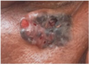
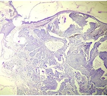
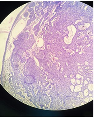
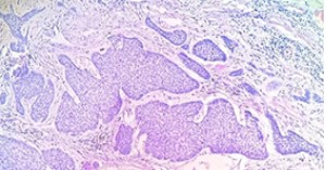
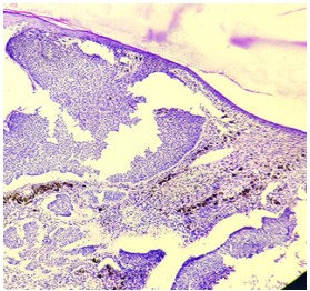
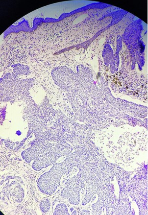
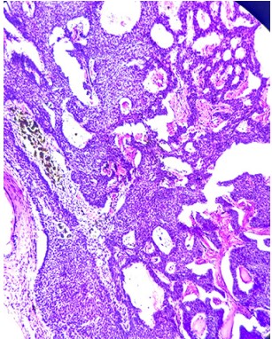
Background: Basal cell carcinomais a slow growing malignant cutaneous non melanocytic tumor that is locally aggressive but it rarely metastasizes. Case of 61 year male presented to the outpatient department with a round, nonhealing ulcer with hyperpigmentation, measuring 2.5x2.1x0.8cm over the right side of face, 1.8 cm below the right eye region for the last 1.5 years. Patient had history of swelling at same site 1.5 year back, as small nodular lesion which was excised. The general physical and systemic examinations were normal. The lesion was surgically excised with wide margins. The specimen on gross measured 2.3x2.1x0.6cm, firm, grey brown to black colored hyperpigmented with rolled out margins and central ulceration measuring 1.6x1.4x0.6cm. Cut section was grey white, nodular with blackish pigmentations. On microscopic examination showed a circumscribed nodular lesion composed of neoplastic cells arranged in large basaloid lobules with peripheral nuclear palisadations. The neoplastic basaloid cells with scant cytoplasm and hyperchromatic nuclei were seen. The stroma was fibromyxoid with focal inflammation. The surface ulceration was noted. The nodules are variable size of more than 0.3cm diameter. Tumor showed epidermal attachment. In areas the reticulate, pseudoglandular pattern of basaloid neoplastic cells were noted with a mucinous stroma. Focal areas of colonization of tumor's complexes with melanocytes and stromal melanophages pigmentation was noted. The tumor showed 0.5cm depth of invasion. The mitotic activity was low, minimal atypia and apoptosis were noted. The lymphovascular, perineural invasion was absent. All peripheral and deep surgical margins were free from tumor. On histopthological findings reported as Basal cell carcinoma-nodular, adenoid cystic and pigmented: mixed histological subtype over face. The patient was kept on regular follow up. There was no evidence of recurrence or any metastasis. Conclusion: We present this rare case of Basal cell carcinoma-nodular, adenoid cystic and pigmented: mixed histological subtype for its clinical and morphological findings. The pathological diagnosis and classification of BCC are essential for the evaluation of the tumour type, its biological behaviour, risk assessment of the recurrence and treatment.
References
- 1. Cameron MC, Lee E, Hibler BP, Barker CA, Mori S, Cordova M, Nehal KS, Rossi AM. Basal cell carcinoma: Epidemiology; pathophysiology; clinical and histological subtypes; and disease associations. J Am Acad Dermatol. 2019; 80(2):303-317.
- 2. Marzuka AG, Book SE. Basal cell carcinoma: Pathogenesis, epidemiology, clinical features, diagnosis, histopathology, and management. Yale J Biol Med 2015; 88:167–179.
- 3. Dubas LE, Ingraffea A.”Nonmelanoma skin cancer”. Facial Plastic Surgery Clinics of North America.2013; 21 (1): 43–53.
- 4. Rogers HW, Weinstock MA, Feldman SR, Coldiron BM. Incidence Estimate of Nonmelanoma Skin Cancer (Keratinocyte Carcinomas) in the U.S. Population, 2012. JAMA Dermatol. 2015; 151(10):1081-6.Top of Form
- 5. Jacob A. Observations respecting an ulcer of peculiar character, which attacks the eyelids and other parts of the face. Dublin Hosp Rep Commun Med Surg. 1827; 4:232–9.
- 6. Anthony Wu, Laub Donald. Metastatic basal cell carcinoma: a case report and review of the literature. Eplasty. 2011;11:ic8.
- 7. Clark CM, Furniss M, Mackay-Wiggan JM. Basal cell carcinoma: an evidence-based treatment update. Am J Clin Dermatol 2014;15:197-216.
- 8. Kiiski V, de Vries E, Flohil SC, Bijl MJ, Hofman A, et al. Risk factors for single and multiple basal cell carcinomas. Arch Dermatol 2010; 146: 848–855.
- 9. Nakayama M, Tabuchi K, Nakamura Y, Hara A. Basal cell carcinoma of the head and neck. J Skin Cancer 2011; 2011:496910.
- 10. Jagtap SV, Kumbhar S, Mane A, Kaur P, Wingkar C.Basal cell carcinoma of eyelid. Arch Cytol Histopathol Res. 2019;4(1):102-104.
- 11. Crowson A.N. Basal cell carcinoma: Biology, morphology and clinical implications. Mod. Pathol. 2006;19((Suppl. S19)):S127–S147.
- 12. Wade TR, Ackerman AB. The many faces of basal cell carcinoma. J Dermatol Surg Oncol 1978; 4: 23–28.
- 13. Bartoš V, Kullová M. Basal cell carcinoma of the skin with mixed histomorphology: a comparative study. Cesk Patol. 2016;52(4):222-226.
- 14. Elder D.E., Massi D., Scolyer R.W.R. Skin Tumours. Pathology and Genetics. Volume 11 IARC Press; Lyon, France: 2018. WHO Classification of Skin Tumours.
- 15. Scolyer R.A. Keratinocytic/Epidermal Tumours. In: Elder D.E., Massi D., Scolyer R.A., Willemze R., editors. WHO Classification of Skin Tumours. Volume 11. IACR Publications; Lyon, France: 2018. pp. 23–63.
- 16. Bastiens MT, Hoefnagel J. Differences in age, site distribution and sex between nodular and superficial basal cell carcinomas indicate different types of tumors J Invest Dermatol. 1998;110:880–84.
- 17. Weedon D, Strutton G. Skin pathology. Edinburgh: Churchill Livingstone, 2002: 765–772.
- 18. Compton LA, Murphy GF, Lian CG. Diagnostic immunohistochemistry in cutaneous Neoplasia: An update. Dermatopathology (Basel). 2015;2:15-42.
- 19. Hutchin ME, et al. Sustained Hedgehog signaling is required for basal cell carcinoma proliferation and survival: conditional skin tumorigenesis recapitulates the hair growth cycle. Genes Dev. 2005;19:214–223.
- 20. Ramdial PK, Madaree A, Reddy R, Chetty R. bcl-2 protein expression in aggressive and nonaggressive basal cell carcinomas. J Cutan Pathol. 2000 ;27(6):283-91.
- 21. Silverman MK, Kopf AW, Bart RS, Grin CM, Levenstein MS. Recurrence rates of treated basal cell carcinomas. Part 3: surgical excision. J Dermatol Surg Oncol. 1992;18:471–6.
Data Sharing Statement
There are no additional data available. All raw data and code are available upon request.
Funding
This research received no funding.
Author Contributions
All authors contributed significantly to the work and approve its publication.
Ethics Declaration
This article does not involve any human or animal subjects, and therefore does not require ethics approval.
Acknowledgements
We would like to express our gratitude to the patients, their families, and all those who have contributed to this study.
Conflicts of Interest
No conflicts of interest in this work.
About this article

Cite this article
Sunil V Jagtap, Shubham S Jagtap, Ramnik Singh et al. Basal Cell Carcinoma Mixed Histological Subtype: case report and Review of Literature. Ind Jr of Path: Res and Practice 2024;13(2)61-66.
Licence:
Attribution-Non-commercial 4.0 International (CC BY-NC 4.0)This license
enables reusers to distribute, remix, adapt, and build upon the material in any
medium or format for noncommercial purposes only, and only so long as
attribution is given to the creator.
| Received | Accepted | Published |
|---|---|---|
| February 13, 2024 | May 06, 2024 | June 30, 2024 |
DOI: https://doi.org/10.21088/ijprp.2278.148X.13224.3
Keywords
Basal cell carcinomaAdenoidPigmented BBCMixed histological subtypeSearch for Similar Articles
Similar Articles
- Clinico-Pathological and Immunohistochemical Study of Primary CNS Tumors in a Te...
- The Role of SGLT2 Inhibitors in Cardiovascular Protection Beyond Diabetes: A Co...
- Epidermal Inclusion Cyst Presenting as 4th Ventricle Space Occupying Lesion: A R...
- Silent Retroperitoneal Seminoma Arising from an Occult Undescended Testis: Diagn...
- Scapular Metastasis of Oral Squamous Cell Carcinoma: A Case Report of an Unusual...
Article Level Metrics
Last UpdatedTuesday 22 September 2026, 05:19:01 (IST)
3671
Accesses
12
541
00
Citations
NA
NA
NA
Download citation
Article Keywords
Keyword Highlighting
Highlight selected keywords in the article text.
Timeline
| Received | February 13, 2024 |
| Accepted | May 06, 2024 |
| Published | June 30, 2024 |
licence
Attribution-Non-commercial 4.0 International (CC BY-NC 4.0)
This license
enables reusers to distribute, remix, adapt, and build upon the material in any
medium or format for noncommercial purposes only, and only so long as
attribution is given to the creator.