Full Text (PDF)
Indian Journal of Anatomy 9(3):p 185-189, July – September 2020. | DOI: http://dx.doi.org/10.21088/ija.2320.0022.9320.1
Original Article
Age Related Changes in Lateral Ventricles of Brain in Normal Individuals by CT Method
Sharada B Menasinkai, Prof & HOD, Department of Anatomy, Adichunchanagiri University (ACU), Karnataka 571418, India. , Mallika B1 , Sharada B Menasinkai2 , Brahmendra M3
Author Information
Licence:
Indian Journal of Anatomy 9(3):p 185-189, July – September 2020. | DOI: http://dx.doi.org/10.21088/ija.2320.0022.9320.1
How Cite This Article:
Mallika B, Sharada B Menasinkai, Brahmendra M. Age Related Changes in Lateral Ventricles of Brain in Normal Individuals by CT Method. Indian J Anat. 2020;9(3):185–189.
Timeline
Received : N/A
Accepted : N/A
Published : N/A
Abstract
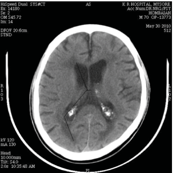
Context: As human brain ages, characteristic structural changes occur that are considered to be normal. Knowledge of age related changes is important before analysing the abnormal findings. As age advances there is regression of brain tissue and thus ventricles size enlarges.
Aims: To analyse the morphometric measurements of lateral ventricles of the brain in different age group in both genders. Material and Methods: The study was done from Feb 2009 to July 2010. Data was collected from the CT scans Dept of Radiology Mysore Medical College & Research Institute. 200 normal CT scans (100 male, 100 female) in the age group of 10–80 yrs were collected.
Results: Right frontal horn measures 28.8 ± 2.5 in youngest and 30.3 ± 2.5 in oldest group. Right body part measures 47.9 ± 2.9 in youngest and 49.0 ± 1.9 in oldest group. Posterior horn measures 26.2 ± 1.9 in youngest age group and 26.0 ± 1.4 in oldest age group. Inferior horns could not be visible in few scans and were taken wherever possible as transverse diameter (TD) and vertical diameter (VD). All the measurements were found to be more in age group > 60 yrs compared to younger age group. Statistical methods: Data was analysed by using SPSS 2004 version, Standard Deviation and Independent ‘t’ test, analysis of variant and P – value.
Conclusion: Advances in CT imaging helps us to understand the normal structure and function of brain. The present study has shown increase in ventricular measurements as age advances.
References
No records found.
About this article
Cite this article
Mallika B, Sharada B Menasinkai, Brahmendra M. Age Related Changes in Lateral Ventricles of Brain in Normal Individuals by CT Method. Indian J Anat. 2020;9(3):185–189.
Licence:
| Received | Accepted | Published |
|---|---|---|
| N/A | N/A | N/A |
DOI: http://dx.doi.org/10.21088/ija.2320.0022.9320.1
Keywords
CT brain; Lateral Ventricles of brain; brain atrophySearch for Similar Articles
Similar Articles
- Nasal Indices of Two Regions of Southern Rajasthan: A Cross-Sectional Study
- Anatomic Variation of the Hepatic Artery and its Importance in Preoperative Pla...
- Unveiling the Coracoulnaris: A Rare Variant of the Coracobrachialis Muscle: Cada...
- Effect of Chemotherapy on Nerve Conduction Velocity
- Morphology of The Liver and Its Sulci in The Senegalese Melanoderm
Article Level Metrics
Last UpdatedMonday 22 June 2026, 04:46:14 (IST)
2341
Accesses
0
400
00
Citations
NA
NA
NA
Download citation
Article Keywords
Keyword Highlighting
Highlight selected keywords in the article text.
Timeline
| Received | N/A |
| Accepted | N/A |
| Published | N/A |