Full Text (PDF)
Indian Journal of Anatomy 15(1):p 19-22, Jan - April 2026. | DOI: https://doi.org/10.21088/ija.2320.0022.15126.3
Original Article
Unveiling the Coracoulnaris: A Rare Variant of the Coracobrachialis Muscle: Cadaveric Observation with Embryological and Clinical Correlation
Soumitra Trivedi, Udayan Sastri, Surbhi Singh
Author Information
Licence:
Attribution-Non-commercial 4.0 International (CC BY-NC 4.0)This license enables reusers to distribute, remix, adapt, and build upon the material in any medium or format for noncommercial purposes only, and only so long as attribution is given to the creator.
Indian Journal of Anatomy 15(1):p 19-22, Jan - April 2026. | DOI: https://doi.org/10.21088/ija.2320.0022.15126.3
How Cite This Article:
Sastri U, Singh S, Trivedi S. Unveiling the Coracoulnaris: A Rare Variant of the Coracobrachialis Muscle: Cadaveric Observation with Embryological and Clinical Correlation. Indian J Anat. 2026;15(1):19-22.Timeline
Received : January 12, 2026
Accepted : February 16, 2026
Published : May 30, 2026
Abstract
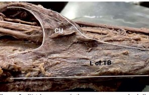
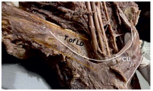
Purpose: The coracobrachialis muscle shows considerable morphological variability, some forms of which may have important surgical and radiological implications. We describe a rare variant in which the coracobrachialis continues distally as an accessory muscle belly coracoulnaris crossing the neurovascular bundle and inserting into the olecranon process. Methods: The variation was identified during routine cadaveric dissection of an embalmed male cadaver aged approximately 70 years. Detailed anatomical dissection was performed to document the origin, course, insertion, and relationships of the variant muscle. Results: The coracobrachialis originated normally from the apex of the coracoid process but did not insert into the humeral shaft. Instead, it formed an elongated accessory belly that crossed superficial to the median nerve and brachial vessels, fused with the long head of the triceps brachii, and inserted into the olecranon process. An additional anomalous attachment to the tendon of latissimus dorsi was observed. Conclusion: This rare configuration represents persistence of embryonic muscle primordia and may predispose to neurovascular compression or complicate surgical and radiological procedures involving the arm and axilla. Awareness of such variants is essential for clinicians working in this region.
References
- 1. Standring S (2020) Gray’s anatomy for students, 4th edn. Elsevier, London.
- 2. Piagkou M, Tsakotos G, Triantafyllou G, Koutserimpas C, Chytas D, Karampelias V, Pantekidis I, Triantafyllou A, Natsis K (2023) Coracobrachialis muscle morphology and coexisted neural variants: a cadaveric case series. Surg Radiol Anat 45:1117–1124.
- 3. Parekh RP, Sawant SP, Rizvi S (2017) Third head of coracobrachialis muscle. Int J Adv Case Rep 4:148–150.
- 4. Vollala VR, Nagabhooshana S, Bhat SM, Potu BK, Rakesh V (2008) Multiple accessory structures in the upper limb of a single cadaver. Singapore Med J 49:959–961.
- 5. Olewnik Ł, Paulsen F, Tubbs RS, Zielińska N, Szewczyk B, Karauda P, Polguj M (2020) Potential compression of the musculocutaneous, median and ulnar nerves by a very rare variant of the coracobrachialis longus muscle. Folia Morphol 80:707–713.
- 6. Tsakotos G, Triantafyllou G, Olewnik Ł, Georgiev GP, Koutserimpas C, Karampelias V, Zielińska N, Piagkou M (2023) A bilateral symmetric accessory coracobrachialis muscle combined with an interconnection of the musculocutaneous nerve with the median nerve. Cureus 15:e36412. 7. Kopuz C, Icten N, Yildirim M (2003) A rare accessory coracobrachialis muscle: a review of the literature. Surg Radiol Anat 24:406–410.
- 8. Jiang LS, Cui YM, Zhou ZD, Dai LY (2007) Stabilizing effect of the transferred conjoined tendon on shoulder stability. Knee Surg Sports Traumatol Arthrosc 15:800–805.
- 9. Wood J (1867) On human muscular variations and their relation to comparative anatomy. J Anat Physiol 1:44–59.
- 10. Lewis WH (1910) The development of the muscular system. In: Keibel F, Mall FP (eds) Manual of embryology, vol 2. JB Lippincott, Philadelphia, pp 455–522.
Data Sharing Statement
There are no additional data available. All raw data and code are available upon request.
Funding
This research received no funding.
Author Contributions
All authors contributed significantly to the work and approve its publication.
Ethics Declaration
Provide information related to the Ethics Committee approval with approval number OR write This article does not involve any human or animal subjects, and therefore does not require ethics approval.
Acknowledgements
We would like to express our gratitude to the patients, their families, and all those who have contributed to this study.
Conflicts of Interest
The authors report no conflicts of interest in this work.
About this article
Cite this article
Sastri U, Singh S, Trivedi S. Unveiling the Coracoulnaris: A Rare Variant of the Coracobrachialis Muscle: Cadaveric Observation with Embryological and Clinical Correlation. Indian J Anat. 2026;15(1):19-22.
Licence:
Attribution-Non-commercial 4.0 International (CC BY-NC 4.0)This license enables reusers to distribute, remix, adapt, and build upon the material in any medium or format for noncommercial purposes only, and only so long as attribution is given to the creator.
| Received | Accepted | Published |
|---|---|---|
| January 12, 2026 | February 16, 2026 | May 30, 2026 |
DOI: https://doi.org/10.21088/ija.2320.0022.15126.3
Keywords
Soumitra TrivediProfessor & HeadDepartment of AnatomyAll India Institute of Medical SciencesRaipurChhattisgarhIndia. E-mail: dr.somit@aiimsraipur.edu.inSearch for Similar Articles
Similar Articles
- Nasal Indices of Two Regions of Southern Rajasthan: A Cross-Sectional Study
- Anatomic Variation of the Hepatic Artery and its Importance in Preoperative Pla...
- Effect of Chemotherapy on Nerve Conduction Velocity
- Morphology of The Liver and Its Sulci in The Senegalese Melanoderm
- Anatomical Variations in the inferior Neurovascular relationships of the thyroid...
Article Level Metrics
Last UpdatedTuesday 28 July 2026, 16:09:05 (IST)
3048
Accesses
16
424
00
Citations
NA
NA
NA
Download citation
Article Keywords
Keyword Highlighting
Highlight selected keywords in the article text.
Timeline
| Received | January 12, 2026 |
| Accepted | February 16, 2026 |
| Published | May 30, 2026 |
licence
Attribution-Non-commercial 4.0 International (CC BY-NC 4.0)
This license enables reusers to distribute, remix, adapt, and build upon the material in any medium or format for noncommercial purposes only, and only so long as attribution is given to the creator.