Full Text (PDF)
Indian Journal of Forensic Medicine and Pathology 13(3):p 399-403, July – September 2020. | DOI: https://doi.org/10.21088/ijfmp.0974.3383.13320.6
Original Article
Age Estimation from Pulpal Width of Maxillary Central Incisors: A Digital Radiographic Study
Karthikeya Patil, Bharathi Penumatsa1 null, Karthikeya Patil2 null, Mahima V Guledgud3 null, Poornima Chandran4 null
Author Information
Licence:
Attribution-Non-commercial 4.0 International (CC BY-NC 4.0)This license enables reusers to distribute, remix, adapt, and build upon the material in any medium or format for noncommercial purposes only, and only so long as attribution is given to the creator.
Indian Journal of Forensic Medicine and Pathology 13(3):p 399-403, July – September 2020. | DOI: https://doi.org/10.21088/ijfmp.0974.3383.13320.6
How Cite This Article:
Penumatsa B, Patil K, Guledgud MV, et al. Age Estimation from pulpal width of Maxillary Central Incisors – A Digital Radiographic Study. Indian Journal of Forensic Medicine & Pathology. 2020;13(3):399–403.Timeline
Received : July 02, 2020
Accepted : July 20, 2020
Published : August 30, 2020
Abstract
Background: Age is one of the prime factors employed to establish the identity of an individual and the use of teeth for this purpose has been considered reliable. The measurement of reduction in pulpal width with advancing age due to secondary dentin deposition can been used as indicators of age.
Aim: To estimate the age of patients using pulpal width of permanent maxillary central incisors and to assess reliability of estimated age based on pulpal width of permanent maxillary central incisors with that of chronological age.
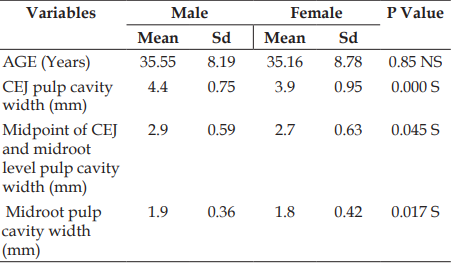
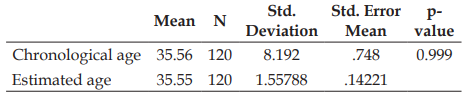
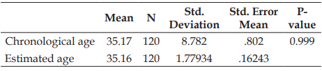
Materials and methods: 240 subjects aged between 20–50 years,with equal gender distribution were included in the study. Digital intraoral periapical radiography (Charge- coupled device sensor) of maxillary central incisors with paralleling angle technique were made for all study subjects and pulpal cavity width was measured at Cemento-enamel junction (CEJ) , midpoint between CEJ and midroot level, midroot level using Planmeca Romexis software. Regression analysis was carried out to obtain the estimated age using pulpal width. Results: The mean estimated age showed no statistical difference from the chronological age in both males and females (p>0.05).
Conclusion: Pulp width serves as a good indicator for age estimation in forensic odontology.
References
- 1. Neville B, Douglas D, Allen CM, Bouquot J. Forensic dentistry. In: Oral and Maxillofacial Pathology. 2nd ed. Philadelphia (PA): W.B. Saunders Co.; 2002. p. 763–83.
- 2. Panchbhai AS. Dental radiographic indicators, a key to age estimation. Dentomaxillofacial Radiology. 2011;40(4):199–212.
- 3. Murray PE, Stanley HR, Mathews JB, Sloan AJ, Smith AJ. Age-related odontometric changes of human teeth. Oral Surgery, Oral Medicine, Oral Pathology, Oral Radiology, and Endodontology. 2002;93(4):474–82.
- 4. Kvaal SI, Kolltveit KM, Thomsen IO, Solheim T. Age estimation of adults from dental radiographs. Forensic Science International. 1995;74(3):175–85.
- 5. Star H, Thevissen P, Jacobs R, Fieuws S, Solheim T, Willems G. Human dental age estimation by calculation of pulp⁄tooth volume ratios yielded on clinically acquired cone beam computed tomography (CBCT) images of monoradicular teeth. Journal of Forensic Sciences. 2011;56(Suppl 1):S77–82.
- 6. Cameriere R, De Luca S, Aleman I, Ferrante L, Cingolani M. Age estimation by pulp/tooth ratio in lower premolars by orthopantomography. Forensic Science International. 2012;214(1–3):105–12.
- 7. Meinl A, Huber CD, Tangl S, Gruber GM, Teschler-Nicola M, Watzek G. Comparison of the validity of three dental methods for the estimation of age at the time of death. Forensic Science International. 2008;178(2-3):96–105.
- 8. Babshet M, Acharya AB, Naikmasur VG. Age estimation in Indians from pulp/tooth area ratio of mandibular canines. Forensic Science International. 2010;197(1–3):125.e1–4.
- 9. Patil SK, Mohankumar KP, Donoghue M. Estimation of age by Kvaal’s technique in sample Indian population to establish the need for local Indian-based formulae. Journal of Forensic Dental Sciences. 2014;6(3):166–70.
- 10. Patil K, Mahima VG, Malleshi NS. Maxillary lateral incisor as biomarker of age-An in vivo radiographic study. Indian Journal of Forensic Odontology. 2009;2:17–20.
- 11. Karjodkar FR. Textbook of Dental and Maxillofacial Radiology. 2nd ed. New Delhi: Jaypee Brothers Medical Publishers; 2009.
- 12. White SC, Pharoah MJ. Oral Radiology: Principles and Interpretation. 6th ed. St. Louis (MO): Mosby; 2009.
- 13. Bodrumlu E, Cicek E, Dundar C. Age and sex-related differences of pulp chamber size in mandibular second molars. Indian Journal of Dental Research. 2013;24(6):742–4.
- 14. Chandler NP, Pitt Ford TR, Monteith BD. Coronal pulp size in molars: A study of bitewing radiographs. International Endodontic Journal. 2003;36(11):757–63.
- 15. Du C, Zhu Y, Hong L. Age-related changes in pulp cavity of incisors as a determinant for forensic age identification. Journal of Forensic Sciences. 2011;56(Suppl 1):S72–6.
- 16. Ginjupally U, Pachigolla R, Sankaran S, Balla S, Pattipati S, Chennoju SK. Assessment of age based on the pulp cavity width of the maxillary central incisors. Journal of Indian Academy of Oral Medicine and Radiology. 2014;26(1):46–9.
- 17. Zaher JF, Fawzy IA, Habibi SR, Ali MM. Age estimation from pulp/tooth area ratio in maxillary incisors among Egyptians using dental radiographic images. Journal of Forensic and Legal Medicine. 2011;18(2):62–5.
Data Sharing Statement
There are no additional data available. All raw data and code are available upon request.
Funding
This research received no funding.
Author Contributions
Whether all authors contributed significantly to the work and approve its publication.
Ethics Declaration
This article does not involve any human or animal subjects, and therefore does not require ethics approval.
Acknowledgements
We would like to express our gratitude to the patients, their families, and all those who have contributed to this study.
Conflicts of Interest
The authors report no conflicts of interest in this work.
About this article

Cite this article
Penumatsa B, Patil K, Guledgud MV, et al. Age Estimation from pulpal width of Maxillary Central Incisors – A Digital Radiographic Study. Indian Journal of Forensic Medicine & Pathology. 2020;13(3):399–403.
Licence:
Attribution-Non-commercial 4.0 International (CC BY-NC 4.0)This license enables reusers to distribute, remix, adapt, and build upon the material in any medium or format for noncommercial purposes only, and only so long as attribution is given to the creator.
| Received | Accepted | Published |
|---|---|---|
| July 02, 2020 | July 20, 2020 | August 30, 2020 |
DOI: https://doi.org/10.21088/ijfmp.0974.3383.13320.6
Keywords
Age estimationchronological ageMaxillary Central Incisorspulpal widthdigital radiographySearch for Similar Articles
Similar Articles
- Diatom Analysis in Thermophilic water bodies: A Study on Hot Water Springs of Ma...
- Morphometric Study of Supra-meatal Triangle and Mastoid process in the Adult dry...
- Histopathological Spectrum of Autopsy Cases: A One Year Retrospective Observatio...
- Forensic Differentiation of Manner of Death in Railway Fatalities: A FourCase Se...
- Understanding assault Patterns: A Cross-sectional study of Cases at Chri, Kelamb...
Article Level Metrics
Last UpdatedSunday 18 October 2026, 12:23:44 (IST)
16262
Accesses
13
3205
00
Citations
NA
NA
NA
Download citation
Article Keywords
Keyword Highlighting
Highlight selected keywords in the article text.
Timeline
| Received | July 02, 2020 |
| Accepted | July 20, 2020 |
| Published | August 30, 2020 |
licence
Attribution-Non-commercial 4.0 International (CC BY-NC 4.0)
This license enables reusers to distribute, remix, adapt, and build upon the material in any medium or format for noncommercial purposes only, and only so long as attribution is given to the creator.