Full Text (PDF)
Indian Journal of Pathology: Research and Practice 14(1):p 31-34, Jan -April 2025. | DOI: https://doi.org/10.21088/ijprp.2278.148X.14125.5
Case Report
A Rare Case Report of Primary Skin-Limited Leucocytoclastic Vasculitis: A Diagnostic and Therapeutic Challenge
Zeeba S. Jairajpuri, Monal Trisal, Shaan Khetrapal, Nida Jalal, Jyoti Mishra
Author Information
Licence:
Attribution-Non-commercial 4.0 International (CC BY-NC 4.0)This license
enables reusers to distribute, remix, adapt, and build upon the material in any
medium or format for noncommercial purposes only, and only so long as
attribution is given to the creator.
Indian Journal of Pathology: Research and Practice 14(1):p 31-34, Jan -April 2025. | DOI: https://doi.org/10.21088/ijprp.2278.148X.14125.5
How Cite This Article:
Monal Trisal, Zeeba S. Jairajpuri, Shaan Khetrapal et al., A Rare Case Report of Primary Skin-Limited Leucocytoclastic Vasculitis: A Diagnostic and Therapeutic Challenge. Ind Jr of Path: Res and Practice 2025;14(1) 31-34.Timeline
Received : February 10, 2025
Accepted : March 05, 2025
Published : April 30, 2025
Abstract
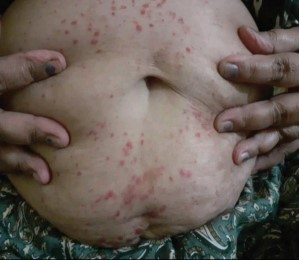
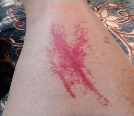
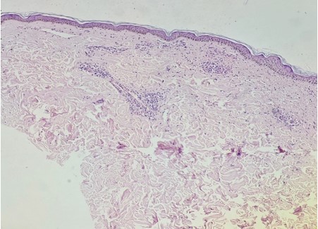
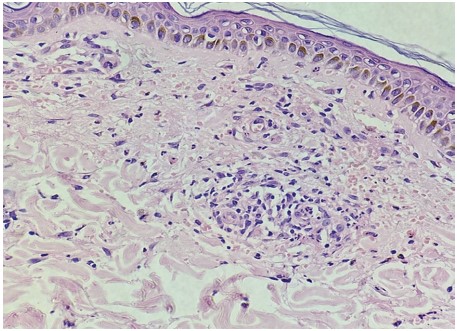
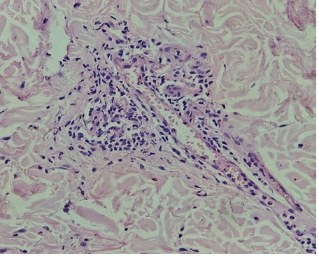
Leucocytoclastic vasculitis (LCV) is a histopathologic term referring to small vessel vasculitis characterized by the infiltration of neutrophils and immune complex deposition. Clinically, LCV presents with palpable purpura, often involving the lower extremities, and may be associated with systemic involvement. We hereby report a rare case of LCV in a 56 year female, highlighting clinical presentation and management strategies for the subset of LCV that remains confined to the skin without systemic manifestations posing diagnostic challenges.
References
- 1. Carlson JA, Chen KR. “Cutaneous vasculitis update: Diagnostic criteria, classification, epidemiology, etiology, pathogenesis, and therapy. ” Am J Dermatopathol. 2006;28(5): 364-383.
- 2. Sunderkotter CH, Zelger B, Chen KR, et al. “Nomenclature of cutaneous vasculitis: A consensus statement from the Dermatology Section of the European Vasculitis Society. ” J Am Acad Dermatol. 2018;79(3): 378-385.
- 3. Poterucha TJ, Wetter DA, Gibson LE, Camilleri MJ. “Microscopic polyangiitis and cutaneous leukocytoclastic vasculitis: A clinicopathologic study of 10 cases. ” J Am Acad Dermatol. 2012;67(5): 945-952.
- 4. Strubchevska K, Kozyk M, Balanescu DV. A Case Report of Leukocytoclastic Vasculitis: Diagnostic Approach and Treatment. Am J Case Rep. 2023:30;24e939541.
- 5. Sakkab R, Fabrikant JM. Leukocytoclastic Vasculitis of the Foot and Ankle: A Case Report With Over Five-Year Follow-Up. Cureus. 2022 May 26;14(5):e25371
- 6. García-Porrua C, González-Gay MA. “Comparative clinical and epidemiological study of hypersensitivity vasculitis versus Henoch-Schönlein purpura in adults. ” Semin Arthritis Rheum. 1999;28(6): 404-412.
Data Sharing Statement
There are no additional data available. All raw data and code are available upon request.
Funding
This research received no funding.
Author Contributions
All authors contributed significantly to the work and approve its publication.
Ethics Declaration
This article does not involve any human or animal subjects, and therefore does not require ethics approval.
Acknowledgements
We would like to express our gratitude to the patients, their families, and all those who have contributed to this study.
Conflicts of Interest
No conflicts of interest in this work.
About this article

Cite this article
Monal Trisal, Zeeba S. Jairajpuri, Shaan Khetrapal et al., A Rare Case Report of Primary Skin-Limited Leucocytoclastic Vasculitis: A Diagnostic and Therapeutic Challenge. Ind Jr of Path: Res and Practice 2025;14(1) 31-34.
Licence:
Attribution-Non-commercial 4.0 International (CC BY-NC 4.0)This license
enables reusers to distribute, remix, adapt, and build upon the material in any
medium or format for noncommercial purposes only, and only so long as
attribution is given to the creator.
| Received | Accepted | Published |
|---|---|---|
| February 10, 2025 | March 05, 2025 | April 30, 2025 |
DOI: https://doi.org/10.21088/ijprp.2278.148X.14125.5
Keywords
Leucocytoclastic vasculitisSmall vessel vasculitisImmune complexPalpablepurpuraSearch for Similar Articles
Similar Articles
- Clinico-Pathological and Immunohistochemical Study of Primary CNS Tumors in a Te...
- The Role of SGLT2 Inhibitors in Cardiovascular Protection Beyond Diabetes: A Co...
- Epidermal Inclusion Cyst Presenting as 4th Ventricle Space Occupying Lesion: A R...
- Silent Retroperitoneal Seminoma Arising from an Occult Undescended Testis: Diagn...
- Scapular Metastasis of Oral Squamous Cell Carcinoma: A Case Report of an Unusual...
Article Level Metrics
Last UpdatedSunday 20 September 2026, 22:00:05 (IST)
3662
Accesses
12
541
00
Citations
NA
NA
NA
Download citation
Article Keywords
Keyword Highlighting
Highlight selected keywords in the article text.
Timeline
| Received | February 10, 2025 |
| Accepted | March 05, 2025 |
| Published | April 30, 2025 |
licence
Attribution-Non-commercial 4.0 International (CC BY-NC 4.0)
This license
enables reusers to distribute, remix, adapt, and build upon the material in any
medium or format for noncommercial purposes only, and only so long as
attribution is given to the creator.