Full Text (PDF)
Indian Journal of Maternal-Fetal & Neonatal Medicine 9(1):p 29-33, january-June 2022. | DOI: https://doi.org/10.21088/ijmfnm.2347.999X.9122.4
Case Report
Huge Lumbosacral Lipomeningomyelocele with Good Post-Natal Surgical Outcome
Dinesh Zinzala, Purvi Desai, Kairavi Desai, Kalpana Kathrotiya, Binodini Chauhan
Author Information
Licence:
Attribution-Non-commercial 4.0 International (CC BY-NC 4.0)This license enables reusers to distribute, remix, adapt, and build upon the material in any medium or format for noncommercial purposes only, and only so long as attribution is given to the creator.
Indian Journal of Maternal-Fetal & Neonatal Medicine 9(1):p 29-33, january-June 2022. | DOI: https://doi.org/10.21088/ijmfnm.2347.999X.9122.4
How Cite This Article:
Purvi Desai, Kairavi desai, Dinesh zinzala et al./Huge Lumbosacral Lipomeningomyelocele with Good Post-Natal Surgical Outcome/Indian Journal of Maternal-Fetal & Neonatal Medicine 2022; 9(1): 29–33.Timeline
Received : October 26, 2021
Accepted : November 10, 2021
Published : June 30, 2022
Abstract
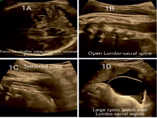
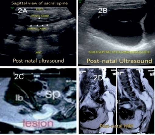
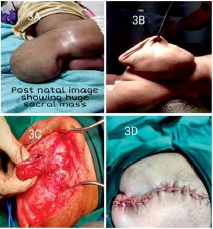
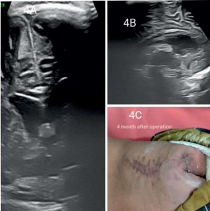
Management of lipomyelomeningocele is one of the most discussed and controversial topics in recent years. To date, there is no consensus on the most appropriate mode of management for lipomyelomeningocele, particularly in asymptomatic patients. It represents a complex disorder that may present with neurological deficit secondary to the inherent tethered cord. Here we present an antenatally diagnosed case of huge lumbosacral lipomeningomyelocele with a good post-natal outcome
References
- 1. McLone DG, Naidich TP.Terminal myelocystocele. Neurosurgery.1985;16:36-43.
- 2. Hoffman HJ, Taecholarn C, Hendrick EB, et al. Management of lipomyelomeningoceles. Experience at the hospital for sick children, Toronto. J Neurosurg. 1985;62:1-8.
- 3. Chocrane DD, Finley C, Kestle J, et al. The patterns of late deterioration in patients with transitional lipomyelomeningocele. Eur J Paedtr Surg.2000;10(suppl 1):13-7.
- 4. Filly RA, Cardoza JD, Goldstein RB, Barkovich AJ: Detection of fetal central nervous system anomalies: a practical level of effort for a routine sonogram. Radiology 172:403–408, 1989.
- 5. Kim SY, McGahan JP, Boggan JE, McGrew W: Prenatal diagnosis of lipomyelomeningocele. J Ultrasound Med 19:801– 805, 2000.
- 6. Kanev PM, Lemire RJ, Loeser JD, Berger MS: Management and long-term follow-up review of children with lipomyelomeningocele, 1952-1987. J Neurosurg 73:48–52, 199.
- 7. Ramamurthi B. Problems in spina bifida in developing countries. J Ind Med Assn.1990;88:6.
Data Sharing Statement
There are no additional data available. All raw data and code are available upon request.
Funding
This research received no funding.
Author Contributions
All authors contributed significantly to the work and approve its publication.
Ethics Declaration
This article does not involve any human or animal subjects, and therefore does not require ethics approval.
Acknowledgements
We would like to express our gratitude to the patients, their families, and all those who have contributed to this study.
Conflicts of Interest
No conflicts of interest in this work.
About this article

Cite this article
Purvi Desai, Kairavi desai, Dinesh zinzala et al./Huge Lumbosacral Lipomeningomyelocele with Good Post-Natal Surgical Outcome/Indian Journal of Maternal-Fetal & Neonatal Medicine 2022; 9(1): 29–33.
Licence:
Attribution-Non-commercial 4.0 International (CC BY-NC 4.0)This license enables reusers to distribute, remix, adapt, and build upon the material in any medium or format for noncommercial purposes only, and only so long as attribution is given to the creator.
| Received | Accepted | Published |
|---|---|---|
| October 26, 2021 | November 10, 2021 | June 30, 2022 |
DOI: https://doi.org/10.21088/ijmfnm.2347.999X.9122.4
Keywords
Lumbosacral lipomeningomyeloceleAsymptomatic patientsUltrasonographySacrococcygeal teratomaSearch for Similar Articles
Similar Articles
- Maternal Mental-Health During Pregnancy and Postpartum Period: A Review of Recen...
- Prenatal Diagnosis of Costello Syndrome: A Case Report
- Innovations in Obstetric Hemorrhage Management: The Evolution of Transvaginal Ba...
- Maternal Plasma Cytokines and the Subsequent Risk of Uterine Atony and Postpartu...
- Amniotic Band Syndrome
Article Level Metrics
Last UpdatedSaturday 17 October 2026, 14:07:35 (IST)
1730
Accesses
28
189
00
Citations
NA
NA
NA
Download citation
Article Keywords
Keyword Highlighting
Highlight selected keywords in the article text.
Timeline
| Received | October 26, 2021 |
| Accepted | November 10, 2021 |
| Published | June 30, 2022 |
licence
Attribution-Non-commercial 4.0 International (CC BY-NC 4.0)
This license enables reusers to distribute, remix, adapt, and build upon the material in any medium or format for noncommercial purposes only, and only so long as attribution is given to the creator.