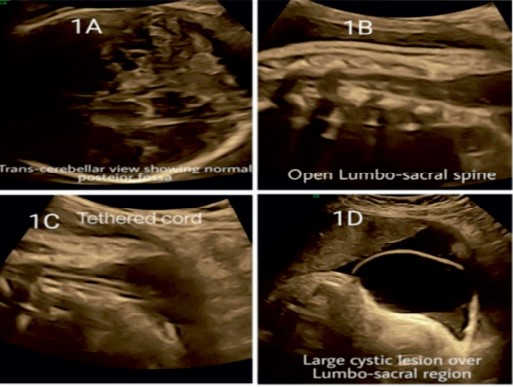
Description: Fig. 1: ultrasound finding at 36 weeks. 1A- normal posterior fossa in trans-cerebellar view; 1B- open lumbo sacral spine in saggital view; 1C - Neural elements extending in the lesion-tethered cord; 1D- large cystic lesion at lumbosacral region.
Heading
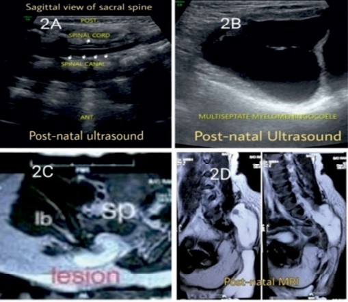
Description: Fig. 2: Showing post natal ultrasound and MRI findings. 2A -Tethered spinal cord, 2B- multiseptate myelomeningocele at lumbosacral region, 2C and 2D -MRI images showing spinal defect with lipomatous tissue covered with skin.
Heading
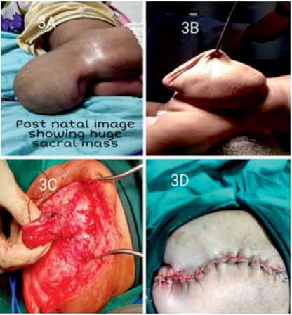
Description: Fig. 3: Showing intra-operative finding.
3A and 3B post natal image of huge lumbosacral mass
3C intraoperative picture
3D postoperative operative site
Heading
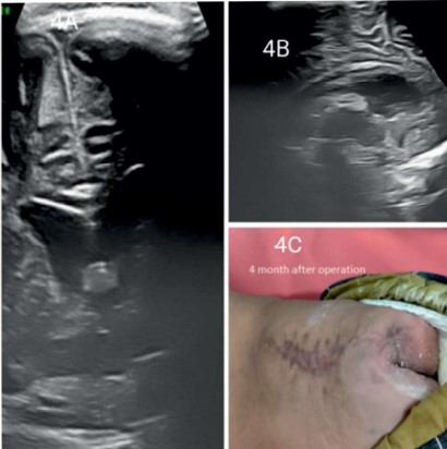
Description: Fig. 4: Showing usg finding and operative site 4 months post surgery.
4A coronal view and 4B sagital view showing normal anterior complex of brain in Fontanalle scan
4C scar site 4 month post surgery