Full Text (PDF)
Indian Journal of Obstetrics and Gynecology 12(3):p 135-137, July-September 2024. | DOI: http://dx.doi.org/10.21088/ijog.2321.1636.12324.5
Case Report
An Incidental Dilemma of Degenerated Stony Parasitic Fibroid: A Rare Case Report
Kothuni Christina, Ananya Das, Yookarin Khonglah, Pranjal Kalita
Author Information
Licence:
Attribution-Non-commercial 4.0 International (CC BY-NC 4.0)This license enables reusers to distribute, remix, adapt, and build upon the material in any medium or format for noncommercial purposes only, and only so long as attribution is given to the creator.
Indian Journal of Obstetrics and Gynecology 12(3):p 135-137, July-September 2024. | DOI: http://dx.doi.org/10.21088/ijog.2321.1636.12324.5
How Cite This Article:
Das A, Chakre KC, Khonglah Y, et al. An incidental dilemma of degenerated stony parasitic fibroid: a rare case report. Indian J Obstet Gynecol. 2024;12(3):135-7.Timeline
Received : May 08, 2024
Accepted : June 09, 2024
Published : June 29, 2024
Abstract
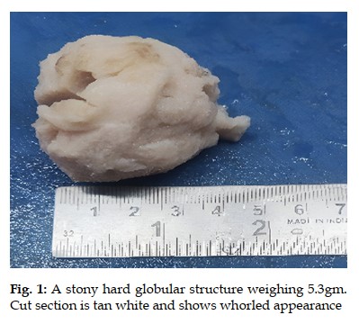
Wandering or parasitic fibroid are rare variant of gynecologic entity and are extrauterine neoplasms occurring in unusual locations where the patient presents atypically or incidentally and at times mislead the radiological findings due to its atypical presentations. Due to the rarity of its presentation and an incidental finding, we present a case of 38 yrs old nulligravida with a myoma of 10 weeks sized, failed medical treatment with an incidental finding of a parasitic degenerated fibroid resembling a stone which was later confirmed as a case of degenerated wandering fibroid.
References
- 1. Epideomology of uterine fibroids: a systemic review. Stewart EA, Cookson CL, Gandolfo RA, Schulze-Rath R. BJOG. 2017;124:1501-1512. [PubMed] [Google Scholar]
- 2. FIGO classification system (PALM-COEIN) for causes of abnormal uterine bleeding in nongravid women of reproductive age. Munro MG, Critchley HO, Broder MS, Fraser IS. Int J Gynaecol Obstet. 2011;113:3-13.
- 3. Huang P, Chang W, Huang S. Iaotrogenic parasitic myoma: A case report and review of literature. Taiwanese Journal of Obstetrics and Gynecology. 53 (2014):392-96.
- 4. Parasitic myoma after morcellation. Sinha R, Sundaram M, Lakhotia S, Kadam P, Rao G, Mahajan C. J GynecolEndosc Surg. –1:113;2009 115.
- 5. Kelly H, Cullen T. Myomata of the Uterus, Philadelphia, 1990. WB Saunders.
- 6. Robin S, Cotran R, Kumar V. Pathologic basis of disease, 3rd edn, Philadelphia, 1984. WB Saunders.
- 7. Najila F, Alampady K, Prasad S, David M. Leiomyoma beyond the Uterus: Unusual locations, rare manifestations. Radiographics. 2008;28:1931-48.
- 8. Vashitha P, Sharma M, Gupta B, Haq M. Wandering Fibroid Presented as Acute Abdomen: A rare case with diagnostic dilemma. J South Asian Feder Obst Gynae 2023;15(4):478-479.
- 9. Gaspere C, Roberta G, Gloria C, Edgardo S. Parasitic myomas after laparoscopic surgery: an emerging complication in the use of morcellator? Description of four cases. Fertil Steril. American Society of Reproductive Medicine. 2011; 96(2):90-6.
Data Sharing Statement
There are no additional data available.
Funding
This research received no funding.
Author Contributions
All authors contributed significantly to the work and approve its publication.
Ethics Declaration
This article does not involve any human or animal subjects, and therefore does not require ethics approval
Acknowledgements
Information Not Provided
Conflicts of Interest
No conflicts of interest in this work.
About this article
Cite this article
Das A, Chakre KC, Khonglah Y, et al. An incidental dilemma of degenerated stony parasitic fibroid: a rare case report. Indian J Obstet Gynecol. 2024;12(3):135-7.
Licence:
Attribution-Non-commercial 4.0 International (CC BY-NC 4.0)This license enables reusers to distribute, remix, adapt, and build upon the material in any medium or format for noncommercial purposes only, and only so long as attribution is given to the creator.
| Received | Accepted | Published |
|---|---|---|
| May 08, 2024 | June 09, 2024 | June 29, 2024 |
DOI: http://dx.doi.org/10.21088/ijog.2321.1636.12324.5
Keywords
Wandering fibroidParasitic fibroidEctopic fibroidIncidental dilemmaDegenerated fibroidSearch for Similar Articles
Similar Articles
- When Eclampsia Strikes the Brain Twice
- Emergency Management of Cesarean Scar Pregnancy: A Rare Case of Life-Threatenin...
- The Efficacy of Structured Teaching Programs in Enhancing Pregnancy Management...
- Audit of Caesarean Sections by the Robson Ten Group Classification: A Six-Month...
- A Cross-Sectional Study on the Epidemiology and Risk Factors of Uterine Fibroids
Article Level Metrics
Last UpdatedThursday 18 June 2026, 04:27:55 (IST)
1422
Accesses
5
152
00
Citations
NA
NA
NA
Download citation
Article Keywords
Keyword Highlighting
Highlight selected keywords in the article text.
Timeline
| Received | May 08, 2024 |
| Accepted | June 09, 2024 |
| Published | June 29, 2024 |
licence
Attribution-Non-commercial 4.0 International (CC BY-NC 4.0)
This license enables reusers to distribute, remix, adapt, and build upon the material in any medium or format for noncommercial purposes only, and only so long as attribution is given to the creator.