Full Text (PDF)
New Indian Journal of Surgery 14(3):p 41-48, July-September 2023. | DOI: https://dx.doi.org/10.21088/nijs.0976.4747.14323.6
Case Report
A Case Series on Hydatid Cyst Disease: Usual and Unusual Sites with Different Approaches and Management
Binoy Kumar Behera, Muruganandam G.M., J.G. Bhatt, J.G. Vagadia
Author Information
Licence:
Attribution-Non-commercial 4.0 International (CC BY-NC 4.0)This license enables reusers to distribute, remix, adapt, and build upon the material in any medium or format for noncommercial purposes only, and only so long as attribution is given to the creator
New Indian Journal of Surgery 14(3):p 41-48, July-September 2023. | DOI: https://dx.doi.org/10.21088/nijs.0976.4747.14323.6
How Cite This Article:
Timeline
Received : May 13, 2023
Accepted : June 30, 2023
Published : September 25, 2023
Abstract
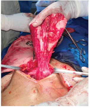
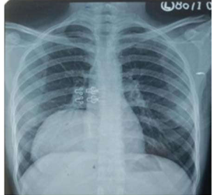
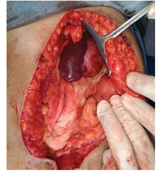
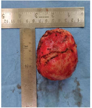
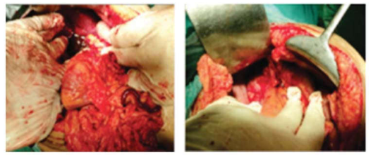
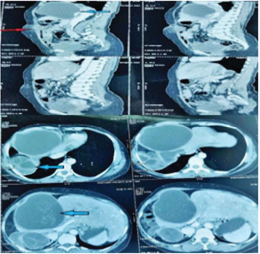
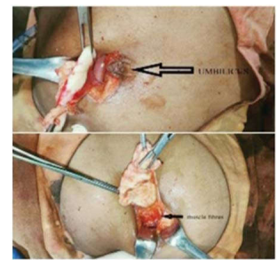
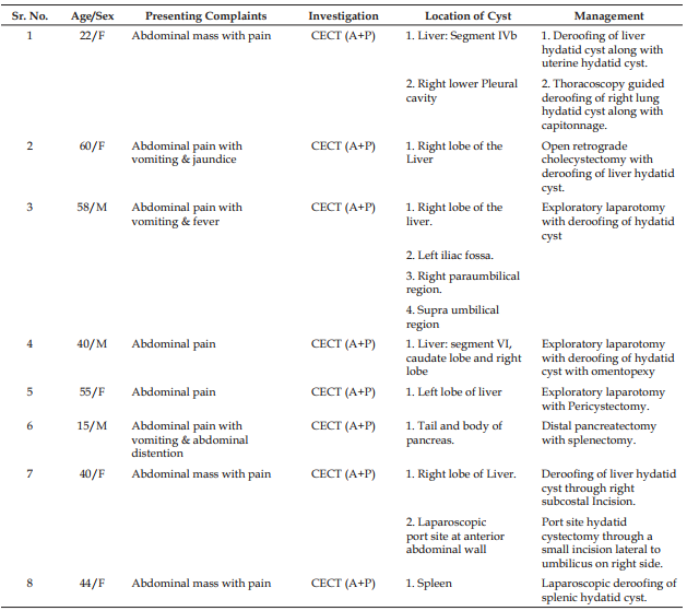
Hydatid disease is a cyclo zoonotic parasitic infection caused by Echinococcus granulosus. This disease is commonly found in the liver and lungs but no organ of the body is immune. When found at unusual sites in the body they show atypical presentations and pose a diagnostic challenge. In such conditions, a high index of suspicion, radiological investigations as well as histopathological examination are necessary in establishing the diagnosis. We present a review of the occurrence of hydatid disease at common as well as unusual sites and our experience with clinical presentation and management of hydatid disease.
References
- 1. Wani RA, Wani I, Malik AA, Parray FQ, Wani AA, Dar AM. Hydatid disease at unusual sites. International Journal of Case Reports and Images 2012;3(6):1–6.
- 2. Kullolli GK, Vaidya MK, Patil R. Unusual Presentations of Hydatid Cyst - A Case Study. Int J Sci Stud 2017;5(1):218-222.
- 3. Zaidi, S.H. Some rare presentations of hydatid cysts: two case reports. Cases Journal 2, 62 (2009). https://doi.org/10.1186/1757-1626-2-62
- 4. Khanna AK, Prasanna GV, Khanna R, Khanna A. Unusual sites of hydatid cysts in India. Trop Doct 2005 Oct;35(4):233–5
- 5. Ozmen MM, Moran M, Karakahya M, Coskun F. Recurrent acute pancreatitis due to a hydatid cyst of the pancreatic head: a case report and review of the literature. JOP J Pancreas (Online) 2005;6:354–8
- 6. Tika Ram Bhandari, Sudha Shahi, “Simultaneous Hydatid Cyst of the Liver and Left Iliac Fossa: An Unusual Case Report”, Case Reports in Surgery, vol. 2019, Article ID 9101425, 4 pages, 2019. https://doi.org/10.1155/2019/9101425
- 7. M. C. Sa oleas, E. P. Misiakos, M. Kouvaraki, M. K. Stamatakos, C. P. Manti, and E. S. Felekouras, “Hydatid disease of Case Reports in Surgery 3 the liver: a continuing surgical problem,“ Archives of surgery, vol. 141, no. 11, pp. 1101–1108, 2006.
Data Sharing Statement
There are no additional data available.
Funding
This research received no funding.
Author Contributions
All authors contributed significantly to the work and approve its publication.
Ethics Declaration
This article does not involve any human or animal subjects, and therefore does not require ethics approval
Acknowledgements
Information Not Provided
Conflicts of Interest
No conflicts of interest in this work
About this article
Cite this article
Licence:
Attribution-Non-commercial 4.0 International (CC BY-NC 4.0)This license enables reusers to distribute, remix, adapt, and build upon the material in any medium or format for noncommercial purposes only, and only so long as attribution is given to the creator
| Received | Accepted | Published |
|---|---|---|
| May 13, 2023 | June 30, 2023 | September 25, 2023 |
DOI: https://dx.doi.org/10.21088/nijs.0976.4747.14323.6
Keywords
Hydatid diseaseUsual sitesUnusual sitesEchinococcus granulosus.Usual sitesUnusual sitesEchinococcus granulosus.Search for Similar Articles
Similar Articles
- Role of Autologous Platelet-Rich Plasma Spray in an Adult with Electrical Burns
- Laparoscopic Management of Large Right Adrenal Teratoma Adherent to Inferior Ven...
- Application of Haemoglobin Spray in Wound Healing in Scald Burns
- Robotic Inguinal Hernia Repair: A Review of Meta-Analyses Comparing Operative Ou...
- A Comparative Study Between Laser Hemorrhoidoplasty and Open Hemorrhoidectomy
Article Level Metrics
Last UpdatedFriday 31 July 2026, 05:01:31 (IST)
3425
Accesses
12
295
00
Citations
NA
NA
NA
Download citation
Article Keywords
Keyword Highlighting
Highlight selected keywords in the article text.
Timeline
| Received | May 13, 2023 |
| Accepted | June 30, 2023 |
| Published | September 25, 2023 |
licence
Attribution-Non-commercial 4.0 International (CC BY-NC 4.0)
This license enables reusers to distribute, remix, adapt, and build upon the material in any medium or format for noncommercial purposes only, and only so long as attribution is given to the creator