Full Text (PDF)
Indian Journal of Dental Education 17(1):p 39-42, January - March 2024. | DOI: https://doi.org/10.21088/ijde.0974.6099.17124.4
Case Report
Warthin’s Tumor in Submandibular Salivary Gland: An Uncommon Extra Parotid Location
Ayushi Kediya, Vishesh Dhawan, Prem Singh
Author Information
Licence:
Attribution-Non-commercial 4.0 International (CC BY-NC 4.0)This license enables reusers to distribute, remix, adapt, and build upon the material in any medium or format for noncommercial purposes only, and only so long as attribution is given to the creator.
Indian Journal of Dental Education 17(1):p 39-42, January - March 2024. | DOI: https://doi.org/10.21088/ijde.0974.6099.17124.4
How Cite This Article:
Ayushi Kediya, Vishesh Dhawan, Prem Singh. Warthin’s Tumor in Submandibular Salivary Gland: An Uncommon Extra Parotid Location. Indian J Dent Educ. 2024;17(1):39- 42.Timeline
Received : January 01, 2024
Accepted : February 20, 2024
Published : March 28, 2024
Abstract
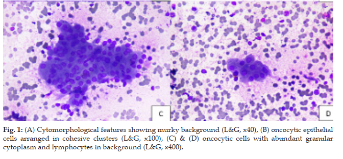
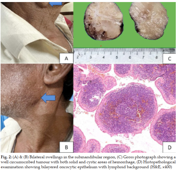
Introduction: Salivary gland tumors are complex to diagnose both on histopathology and cytology. Majority of these tumours arise in the parotid glands, followed by submandibular glands, and rarely in minor salivary glands. Warthin’s tumor (WT), also known as papillary cystadenoma lymphomatosum, is characterized by a dense lymphoid stroma and a double layer of oncocytic epithelium with papillary and cystic architecture. Only 6-10% cases have been reported in the extra-parotid locations i.e. cervical lymph nodes, submandibular gland and larynx. Case Report: Our patient presented with painless bilateral submandibular swellings for 1.5 months with history of chronic smoking of two bundles of biddis per day for 32 years. Conclusion: Hereby, this case was unique in its presentation as it was located in an extra parotid region with bilateral origin, as these locations carry clinical significance in diagnosing and ruling out conditions like lymphoma.
References
- 1. Young A, Okuyemi OT. Malignant salivary gland tumors. In Stat Pearls [Internet] 2023 Jan 12. Stat Pearls Publishing.
- 2. Yankov Y, Todorova K, Stoev L, Dimanov S. Salivary duct carcinoma of the parotid gland: a review article. In Varna Medical Forum 2023 Nov 21 (Vol. 12, No. 2).
- 3. Limaiem F, Jain P. Warthin tumor. In Stat Pearls [Internet] 2023 Jan 1. Stat Pearls Publishing.
- 4. Bajaj P, Garg D, Sabharwal R, Gautam S. Fine Ayushi Kediya, Vishesh Dhawan, Prem Singh. Warthin’s Tumor in Submandibular Salivary Gland: An Uncommon Extra Parotid Location needle aspiration cytology in Warthin’s tumor: a diagnostic tool. Diagnostic Pathology: Open Access. 2015;1(1):102-6.
- 5. Mukunyadzi P. Review of fine-needle aspiration cytology of salivary gland neoplasms, with emphasis on differential diagnosis. Pathology Patterns Reviews. 2002 Dec 1; 118(suppl_1):S100-15.
- 6. Sood N, Borah P. Warthins tumor: Cyto histological spectrum with emphasis on diagnostic difficulties. Diagnostic Cytopathology. 2018 Jul;46(7):613-9.
- 7. Flezar M, Pogacnik A. Warthin's tumour: unusual vs. common morphological findings in fine needle aspiration biopsies. Cytopathology. 2002 Aug; 13(4):232-41.
- 8. Parwani AV, Ali SZ. Diagnostic accuracy and pitfalls in fine needle aspiration interpretation of Warthin tumor. Cancer Cytopathology: Interdisciplinary International Journal of the American Cancer Society. 2003 Jun 25;99(3):166-71.
- 9. Viguer JM, Vicandi B, Jiménez Heffernan JA, López Ferrer P, González Peramato P, Castillo C. Role of fine needle aspiration cytology in the diagnosis and management of Warthin’s tumour of the salivary glands. Cytopathology. 2010 Jun; 21(3):164-9.
- 10. Kim JY, Yoo YS, Kwon JE, Kim HJ, Park K. Fine-needle aspiration cytology with c-kit immunocytochemical staining in the diagnosis of Warthin’s tumor. Acta Cytologica. 2012 Oct 1; 56(5):474-80.
- 11. Chakrabarti I, Basu A, Ghosh N. Oncocytic lesion of parotid gland: A dilemma for cytopathologists. Journal of Cytology/Indian Academy of Cytologists. 2012 Jan;29(1):80.
- 12. K0ybasioglu FF, Onal B, Han U, Adabag A, Sahpaz A. Cytomorphological findings in diagnosis of Warthin tumor. Turkish journal of medical sciences. 2020; 50(1):148-54.
- 13. Yoo GH, Eisele DW, Driben JS, Johns ME, Askin FB. Warthin's tumor: A 40 year experience at the johns hopkins hospital. The Laryngoscope. 1994 Jul; 104(7):799-803.
- 14. Naujoks C, Sproll C, Singh DD, Heikaus S, Depprich R, Kubler NR, Handschel J. Bilateral multifocal Warthin's tumors in upper neck lymph nodes. Report of a case and brief review of the literature. Head & Face Medicine. 2012 Dec;8:1-6.
Data Sharing Statement
There are no additional data available. All raw data and code are available upon request.
Funding
This research received no funding.
Author Contributions
All authors contributed significantly to the work and approve its publication.
Ethics Declaration
This article does not involve any human or animal subjects, and therefore does not require ethics approval.
Acknowledgements
Information not provide.
Conflicts of Interest
The authors report no conflicts of interest in this work.
About this article
Cite this article
Ayushi Kediya, Vishesh Dhawan, Prem Singh. Warthin’s Tumor in Submandibular Salivary Gland: An Uncommon Extra Parotid Location. Indian J Dent Educ. 2024;17(1):39- 42.
Licence:
Attribution-Non-commercial 4.0 International (CC BY-NC 4.0)This license enables reusers to distribute, remix, adapt, and build upon the material in any medium or format for noncommercial purposes only, and only so long as attribution is given to the creator.
| Received | Accepted | Published |
|---|---|---|
| January 01, 2024 | February 20, 2024 | March 28, 2024 |
DOI: https://doi.org/10.21088/ijde.0974.6099.17124.4
Keywords
CytologyFine Needle Aspiration CytologySubmandibular GlandWarthin’s TumorSearch for Similar Articles
Similar Articles
- Post Extraction Surgical Ciliated Cyst of Maxilla: Case Report of Unusual Pathog...
- Natural Tooth Surgical Stent (NTSS) Technique For Prosthodontically Driven Rehab...
- Growth Meets Diagnosis: Revisiting Synchondrosis in the Orthodontic Era
- The Silent Struggle to Breathe: A Review on Obstructive Sleep Apnea
- Evaluating Awareness of Oral-Systemic Health Interactions in Urbanized Indian So...
Article Level Metrics
Last UpdatedThursday 18 June 2026, 02:49:10 (IST)
1364
Accesses
10
435
00
Citations
NA
NA
NA
Download citation
Article Keywords
Keyword Highlighting
Highlight selected keywords in the article text.
Timeline
| Received | January 01, 2024 |
| Accepted | February 20, 2024 |
| Published | March 28, 2024 |
licence
Attribution-Non-commercial 4.0 International (CC BY-NC 4.0)
This license enables reusers to distribute, remix, adapt, and build upon the material in any medium or format for noncommercial purposes only, and only so long as attribution is given to the creator.