Full Text (PDF)
Journal of Cardiovascular Medicine and Surgery 12(1):p 11-20, Jan-April 2026. | DOI: DOI: 10.21088/jcms.2454.7123.12126.2
Review Article
Hemodynamic Monitoring by Point-of-Care Ultrasound: A Multiorgan, Physiology-Driven Approach to Precision Cardiac Critical Care (From Static Assessment to Dynamic Functional Assessment)
Author Information
Licence:
Attribution-Non-commercial 4.0 International (CC BY-NC 4.0)This license enables reusers to distribute, remix, adapt, and build upon the material in any medium or format for noncommercial purposes only, and only so long as attribution is given to the creator.
Journal of Cardiovascular Medicine and Surgery 12(1):p 11-20, Jan-April 2026. | DOI: DOI: 10.21088/jcms.2454.7123.12126.2
How Cite This Article:
Sambhunath Das, Roja Emani. Hemodynamic Monitoring by Point-of-Care Ultrasound: A Multiorgan, Physiology-Driven Approach to Precision Cardiac Critical Care (From Static Assessment to Dynamic Functional Assessment). Indian J Cardiovasc Med Surg. 2026; 12(1): 11–20.Timeline
Received : March 03, 2026
Accepted : April 02, 2026
Published : April 30, 2026
Abstract
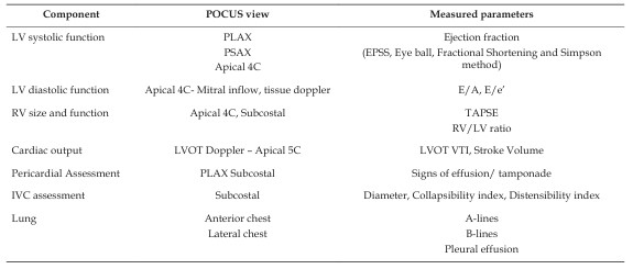
One of the most critical insights required for the management of a critically ill patient is to know the hemodynamic parameters. Over years, hemodynamic monitoring has evolved from simple bedside clinical examination to invasive monitoring techniques such as pulmonary artery catheterization, pulse contour analysis and is advancing into non-invasive methods improving the safety profile of the patient. Traditional invasive techniques such as pulmonary artery catheterization and transpulmonary thermodilution have provided detailed physiological insights but are associated with procedural risks and intermittent data acquisition. Over the past two decades, point-of-care ultrasound (POCUS) has emerged as an advanced bedside monitoring modality enabling real-time, repeatable, and non-invasive hemodynamic assessment. POCUS allows integrated evaluation of cardiac output, ventricular function, preload, afterload, pulmonary congestion, and systemic venous congestion. By facilitating rapid shock phenotyping and guiding individualized resuscitation strategies, it represents a paradigm shift from static pressure-based monitoring toward dynamic, physiology-oriented multiorgan assessment. This review synthesizes current evidence regarding the clinical application of cardiac, vascular, pulmonary, and venous Doppler ultrasound in hemodynamic monitoring, discusses limitations, and outlines future directions in precision cardiac critical care
References
- 1. Noor A, Liu M, Jarman A, Yamanaka T, Kaul M. Point-of-Care Ultrasound Use in Hemodynamic Assessment. Biomedicines. 2025 Jun 10; 13(6). doi:10.3390/biomedicines13061426
- 2. Chanda AH, Idayathulla S, Zulfiqar H, Fatima M, Balal A, Alhussein MMK, et al. POCUS (Point of Care Ultrasound): Hemodynamic Monitoring of Future [Internet]. IntechOpen; 2026 [cited 2026 Feb 28]. Available from: https://www.intechopen.com/onlinef irst/1227661 doi:10.5772/intechopen.1013633
- 3. Naddaf N, Dianati Maleki N, Goldschmidt ME, Kalogeropoulos AP. Point of Care Ultrasound (POCUS) in the Management of Heart Failure: A Narrative Review. J Pers Med. 2024 Jul 18; 14(7): 766. doi:10.3390/jpm14070766 PubMed PMID: 39064020; PubMed Central PMCID: PMC11277924.
- 4. Chengode S. Left ventricular global systolic function assessment by echocardiography. Ann Card Anaesth. 2016 Oct; 19(Supplement): S26–34. doi:10.4103/0971-9784.192617 PubMed PMID: 27762246; PubMed Central PMCID: PMC5100240.
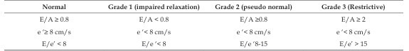
- 5. Greenstein YY, Mayo PH. Evaluation of Left Ventricular Diastolic Function by the Intensivist. CHEST. 2018 Mar 1; 153(3): 723 32. doi:10.1016/j.chest.2017.10.032 PubMed PMID: 29113815.
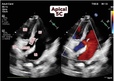
- 6. Mercado P, Maizel J, Beyls C, Titeca-Beauport D, Joris M, Kontar L, et al. Transthoracic echocardiography: an accurate and precise method for estimating cardiac output in the critically ill patient. Crit Care. 2017 Jun 9; 21: 136. doi:10.1186/s13054-017-1737-7 PubMed PMID: 28595621; PubMed Central PMCID: PMC5465531.
- 7. Xie J, Wan J, Xu L, Zhang Y, Chen J. The Accuracy of Velocity-Time Integral Variation and Peak Velocity Variation of the Left Ventricular Outflow Tract in Predicting Fluid Responsiveness in Postoperative Patients Mechanically Ventilated at Low Tidal Volumes. J Cardiothorac Vasc Anesth. 2023 Jun 1; 37(6): 911–8. doi:10.1053/j.jvca.2023.02.009 PubMed PMID: 36931906.
- 8. Monnet X, Shi R, Teboul JL. Prediction of fluid responsiveness. What’s new? Ann Intensive Care. 2022 May 28; 12: 46. doi:10.1186/s13613 022-01022-8 PubMed PMID: 35633423; PubMed Central PMCID: PMC9148319.
- 9. Premkumar M, Kajal K, Gupta A, Izzy M, Roy A, Sihag B, et al. Point-of-care Ultrasound (POCUS)-Guided Pragmatic Fluid and Albumin Resuscitation and Hemodynamic Monitoring in Cirrhosis and Septic Shock. J JCMS / Volume 12 Number 1/January – April 2026 20 Journal of Cardiovascular Medicine and Surgery Clin Exp Hepatol. 2026 Mar 1; 16(2): 103463. doi:10.1016/j.jceh.2025.103463
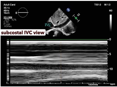
- 10. Ciozda W, Kedan I, Kehl DW, Zimmer R, Khandwalla R, Kimchi A. The efficacy of sonographic measurement of inferior vena cava diameter as an estimate of central venous pressure. Cardiovasc Ultrasound. 2016 Aug 20; 14: 33. doi:10.1186/s12947-016-0076-1 PubMed PMID: 27542597; PubMed Central PMCID: PMC4992235.
- 11. Di Nicolò P, Tavazzi G, Nannoni L, Corradi F. Inferior Vena Cava Ultrasonography for Volume Status Evaluation: An Intriguing Promise Never Fulfilled. J Clin Med. 2023 Mar 13; 12(6): 2217. doi:10.3390/jcm12062217 PubMed PMID: 36983218; PubMed Central PMCID: PMC10053997
- 12. Right Ventricular Apical Contractility in Acute Pulmonary Embolism: The McConnell Sign Revisited - López-Candales - 2010 - Echocardiography - Wiley Online Library [Internet]. [cited 2026 Feb 28]. Available from: https://onlinelibrary.wiley.com/ doi/10.1111/j.1540-8175.2009.01103.x
- 13. Andrei S, Bahr PA, Nguyen M, Bouhemad B, Guinot PG. Prevalence of systemic venous congestion assessed by Venous Excess Ultrasound Grading System (VExUS) and association with acute kidney injury in a general ICU cohort: A prospective multicentric study. Crit Care. 2023 Jun 8; 27: 224. doi:10.1186/ s13054-023-04524-4 PubMed PMID: 37291662; PubMed Central PMCID: PMC10249288.
Data Sharing Statement
There are no additional data available. All raw data and code are available upon request.
Funding
This research received no funding.
Author Contributions
All authors contributed significantly to the work and approve its publication.
Ethics Declaration
This article does not involve any human or animal subjects, and therefore does not require ethics approval.
Acknowledgements
We would like to express our gratitude to the patients, their families, and all those who have contributed to this study.
Conflicts of Interest
The authors report no conflicts of interest in this work.
About this article
Cite this article
Sambhunath Das, Roja Emani. Hemodynamic Monitoring by Point-of-Care Ultrasound: A Multiorgan, Physiology-Driven Approach to Precision Cardiac Critical Care (From Static Assessment to Dynamic Functional Assessment). Indian J Cardiovasc Med Surg. 2026; 12(1): 11–20.
Licence:
Attribution-Non-commercial 4.0 International (CC BY-NC 4.0)This license enables reusers to distribute, remix, adapt, and build upon the material in any medium or format for noncommercial purposes only, and only so long as attribution is given to the creator.
| Received | Accepted | Published |
|---|---|---|
| March 03, 2026 | April 02, 2026 | April 30, 2026 |
DOI: DOI: 10.21088/jcms.2454.7123.12126.2
Keywords
POCUS (point of care ultrasound)Hemodynamic monitoringCardiac critical careNon-invasive cardiac monitoringFluid responsivenessLung ultrasoundVenous congestionSearch for Similar Articles
Similar Articles
- Giant Intra-cardiac Lipoma in the Right Atrium
- Case Report: Neurophysiological Facilitation of Respiration in an Infant with...
- Left Coronary Artery Fistula Masquerading as Coronary Insufficiency: A Rare Sur...
- Aortotomy Closure after Cardiac Valve Replacements: Nightmare for Surgeons
- Rare Neck Angiolipoma Managed with Surgical Excision: A Case Report
Article Level Metrics
Last UpdatedTuesday 28 July 2026, 16:08:57 (IST)
972
Accesses
19
136
00
Citations
NA
NA
NA
Download citation
Article Keywords
Keyword Highlighting
Highlight selected keywords in the article text.
Timeline
| Received | March 03, 2026 |
| Accepted | April 02, 2026 |
| Published | April 30, 2026 |
licence
Attribution-Non-commercial 4.0 International (CC BY-NC 4.0)
This license enables reusers to distribute, remix, adapt, and build upon the material in any medium or format for noncommercial purposes only, and only so long as attribution is given to the creator.