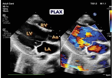
Parasternal long axis view showing right ventricle, left ventricle, left atrium and aorta
Description: No description available.
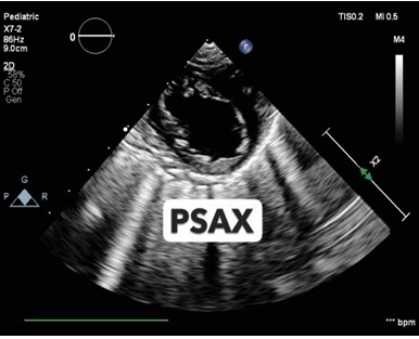
Parasternal short axis view showing left ventricle and right ventricle in short axis. This view is used to visualize for any regional wall motion abnormalities, volume in LV, and estimate LVEF using M-mode
Description: No description available.
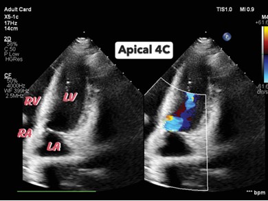
This image shows the apical 4-chamber view which can be used for assessment for LV systolic and diastolic function and RV function
Description: No description available.
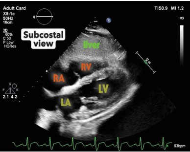
Image showing subcostal view. Most commonly used to interpret RV function and check for any pericardial effusion.
Description: No description available.
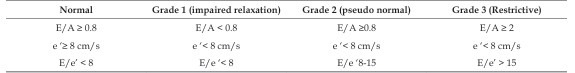
Grading for diastolic dysfunction based on mitral inflow and tissue doppler (adapted from 1)
Description: No description available.
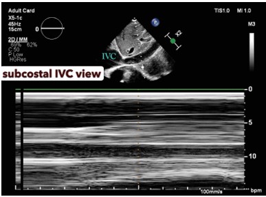
Image showing subcostal IVC view. This view is used to measure IVC diameter and IVC collapsibility or distensibility index using M-mode as shown in the image approximately 2 cms caudal to IVC-RA junction and is used to estimate fluid responsiveness of the patient
Description: No description available.
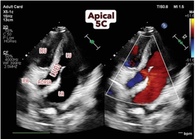
Apical 5 chamber showing Aorta and LVOT in addition to all 4 chambers.This view is used to measure LVOT-VTI using pulse wave doppler for estimation of stroke volume and cardiac output.
Description: No description available.
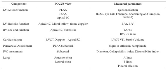
Summary of ultrasonographic views for assessment of heart, lung, IVC