Full Text (PDF)
Ophthalmology and Allied Sciences 12(1):p 27-32, Jan -June 2026. | DOI: DOI: 10.21088/oas.2454.7816.12126.4
Case Report
Familial Foveal Hypoplasia and Review of Literature
Vijay Kumar Maurya, Jagriti Rana, Arti Singh, Anushka Sonkar, Shivani Katheria
Author Information
Licence:
Attribution-Non-commercial 4.0 International (CC BY-NC 4.0)This license enables reusers to distribute, remix, adapt, and build upon the material in any medium or format for noncommercial purposes only, and only so long as attribution is given to the creator.
Ophthalmology and Allied Sciences 12(1):p 27-32, Jan -June 2026. | DOI: DOI: 10.21088/oas.2454.7816.12126.4
How Cite This Article:
Vijay Kumar Maurya, Jagriti Rana, Arti Singh et. al, Familial Foveal Hypoplasia and Review of Literature. Ophthalmol Allied Sci. 2026;12(1): 27–32.Timeline
Received : July 20, 2026
Accepted : August 22, 2026
Published : June 30, 2026
Abstract
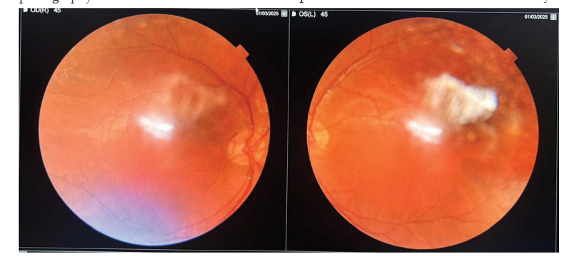
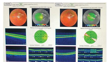
ABSTRACT Objective: To describe a familial presentation of isolated foveal hypoplasia (FH) with detailed clinico-imaging correlation based on fundus photography and spectral-domain optical coherence tomography (SD-OCT), highlighting the diagnostic value of multimodal imaging in this rare condition. Methods: A 22-year-old male proband and his younger sister underwent complete ophthalmic evaluation including best-corrected visual acuity (BCVA), slit-lamp biomicroscopy, dilated fundus examination, Topcon Maestro2 SD-OCT with radial macular scanning protocol, and whole-exome molecular genetic testing. Results: BCVA was 20/200 bilaterally in the proband. Fundus photography revealed absence of the foveal reflex in both eyes, with an incidental chorioretinal lesion in the left eye. SD-OCT demonstrated absent foveal pit with diffuse macular thickening (OD mean 259.9 µm; OS mean 279.6 µm) and persistence of inner retinal layers over the foveal center bilaterally, consistent with Grade 3 FH. A pathogenic FRMD7 gene variant was identified on whole-exome sequencing. His sibling harbored the same variant in heterozygous form. Conclusions: Multimodal imaging – combining fundus photography with SD-OCT macular thickness mapping – provides objective, quantifiable documentation of foveal maldevelopment and enables grading that guides prognosis and counseling in familial FH.
References
- 1. Recchia FM, Carvalho-Recchia CA, Trese MT. Optical coherence tomography in the diagnosis of foveal hypoplasia. Arch Ophthalmol. 2002;120(11):1587-1588.
- 2. Marmor MF, Fulton AB, Holder GE, Miyake Y, Brigell M, Bach M. ISCEV Standard for full-field clinical electroretinography (2008 update). Doc Ophthalmol. 2009;118(1):69-77. doi:10.1007/s10633-008-9155-4.
- 3. Dubis AM, Hansen BR, Cooper RF, Beringer J, Dubra A, Carroll J. Relationship between the foveal avascular zone and foveal pit morphology. Invest Ophthalmol Vis Sci. 2012;53(3):1628-1636. doi:10.1167/iovs.11- 8488.
- 4. Perez Y, Gradstein L, Flusser H, Markus B, Cohen I, Langer Y, Kadir R, Volodarsky M, Halpern GJ, Frayling IM, Sheikh TI, GrossTsur V, Birk OS. Isolated foveal hypoplasia with secondary nystagmus and low vision is associated with a homozygous SLC38A8 mutation. Eur J Hum Genet. 2014;22(6):703- 706. doi:10.1038/ejhg.2013.231.
- 5. Thomas MG, Crosier M, Lindsay S, Kumar A, Araki M, Leroy BP, McLean RJ, Sheth V, Maconachie G, Thomas S, Moore AT, Gottlob I. Abnormal retinal development associated with FRMD7 mutations. Hum Mol Genet. 2014;23(15):4086-4093. doi:10.1093/hmg/ ddu122.
- 6. Tarpey P, Thomas S, Sarvananthan N, Mallya U, Lisgo S, Talbot CJ, Roberts EO, Awan M, Lindsey J, Bussell L, Newall T, Moser P, Alattas S, Leroy BP. Mutations in FRMD7, a newly identified member of the FERM family, cause X-linked idiopathic congenital nystagmus. Nat Genet. 2006;38(11):1242-1244. doi:10.1038/ ng1893.
- 7. Thiadens AA, Somervuo V, van den Born LI, Roosing S, van Schooneveld MJ, Kuijpers RW, van Moll-Ramirez N, Cremers FP, Hoyng CB, Klaver CC. Progressive loss of cones in achromatopsia: an imaging study using spectral-domain optical coherence tomography. Invest Ophthalmol Vis Sci. 2010;51(11):5952-5957. doi:10.1167/iovs. 10-5680.
- 8. Vincent A, Robson AG, Holder GE. Pathognomonic (diagnostic) ERGs. A review and update. Retina. 2013;33(1):5-12. doi:10.1097/IAE.0b013e318279f7f6.
- 9. Thomas MG, Kumar A, Mohammad S, Proudlock FA, Engle EC, Andrews C, Chan WM, Thomas S, Gottlob I. Structural grading of foveal hypoplasia using spectraldomain optical coherence tomography: a predictor of visual acuity? Ophthalmology. 2011;118(8):1653-1660. doi:10.1016/j.ophtha. 2011.01.028.
- 10. Betts-Henderson J, Bartesaghi S, Crosier M, Lindsay S, Chen HL, Salomoni P, Gottlob I, Nicotera P. The nystagmus-associated FRMD7 gene regulates neuronal outgrowth and development. Hum Mol Genet. 2010;19(2):342- 351. doi:10.1093/hmg/ddp500.
- 11. Khan AO. Optics of congenital nystagmus and foveal hypoplasia. Ophthalmology. 2012;119(11):2256-2262. doi:10.1016/j. opht 2012.06.045. Vijay Kumar Maurya, Jagriti Rana, Arti Singh et. al, Familial Foveal Hypoplasia and Review of Literature32 Ophthalmology and Allied Sciences OAS/ Volume 12 Number 1/ January - June 2026
- 12. Gronskov K, Ek J, Brondum-Nielsen K. Oculocutaneous albinism. Orphanet J Rare Dis. 2007;2:43. doi:10.1186/1750-1172-2-43.
- 13. Hingorani M, Hanson I, van Heyningen V. Aniridia. Eur J Hum Genet. 2012;20(10):1011- 1017. doi:10.1038/ejhg.2012.100.
- 14. Lee H, Sheth V, Bibi M, Robson A, Griffiths PG, Abdullah-Suhaimi A, Elshatory Y, Maconachie G, Gottlob I, Thomas MG. . Potential of handheld optical coherence tomography to determine cause of infantile nystagmus in children by using foveal morphology. Ophthalmology. 2013;120(12):2714-2724. doi:10.1016/j. ophtha.2013.07.018.
- 15. McLean RJ, Windridge KC, Gottlob I. Living with nystagmus: a qualitative study. Br J Ophthalmol. 2012;96(7):981-986. doi:10.1136/ bjophthalmol-2011-301519
Data Sharing Statement
There are no additional data available. All raw data and code are available upon request.
Funding
This research received no funding.
Author Contributions
All authors contributed significantly to the work and approve its publication.
Ethics Declaration
This article does not involve any human or animal subjects, and therefore does not require ethics approval.
Acknowledgements
We would like to express our gratitude to the patients, their families, and all those who have contributed to this study.
Conflicts of Interest
No conflicts of interest in this work.
About this article

Cite this article
Vijay Kumar Maurya, Jagriti Rana, Arti Singh et. al, Familial Foveal Hypoplasia and Review of Literature. Ophthalmol Allied Sci. 2026;12(1): 27–32.
Licence:
Attribution-Non-commercial 4.0 International (CC BY-NC 4.0)This license enables reusers to distribute, remix, adapt, and build upon the material in any medium or format for noncommercial purposes only, and only so long as attribution is given to the creator.
| Received | Accepted | Published |
|---|---|---|
| July 20, 2026 | August 22, 2026 | June 30, 2026 |
DOI: DOI: 10.21088/oas.2454.7816.12126.4
Keywords
Foveal HypoplasiaFrmd7 GeneSD-OCTMacular ThicknessFundus PhotographyFamilialX-Linked NystagmusSearch for Similar Articles
Similar Articles
Article Level Metrics
Last UpdatedSaturday 24 October 2026, 19:03:36 (IST)
1905
Accesses
15
157
00
Citations
NA
NA
NA
Download citation
Article Keywords
Keyword Highlighting
Highlight selected keywords in the article text.
Timeline
| Received | July 20, 2026 |
| Accepted | August 22, 2026 |
| Published | June 30, 2026 |
licence
Attribution-Non-commercial 4.0 International (CC BY-NC 4.0)
This license enables reusers to distribute, remix, adapt, and build upon the material in any medium or format for noncommercial purposes only, and only so long as attribution is given to the creator.