Full Text (PDF)
International Journal of Neurology and Neurosurgery 18(2):p 68-72, May-August 2026. | DOI: https://doi.org/10.21088/ijnns.0975.0223.18226.4
Case Report
Cerebellar Tuberculoma Mimicking Ganglioglioma in a Pediatric Patient: A Case Reporting Discordance of Pathology-Radiological Imaging
Shallini Mittal, Rashmi Bhardwaj, Nikita Jajodia, Shibani Saluja, Krishan Kumar Yadav
Author Information
Licence:
Attribution-Non-commercial 4.0 International (CC BY-NC 4.0)This license enables reusers to distribute, remix, adapt, and build upon the material in any medium or format for noncommercial purposes only, and only so long as attribution is given to the creator.
International Journal of Neurology and Neurosurgery 18(2):p 68-72, May-August 2026. | DOI: https://doi.org/10.21088/ijnns.0975.0223.18226.4
How Cite This Article:
Shallini Mittal, Rashmi Bhardwaj, Nikita Jajodia et. al, Cerebellar Tuberculoma mimicking Ganglioglioma in a pediatric patient: A Case reporting discordance of pathology-radiological imaging. Int. J Neuro Neurosurgery. 2026; 18(2): 68-72.Timeline
Received : May 25, 2026
Accepted : July 30, 2026
Published : August 30, 2026
Abstract
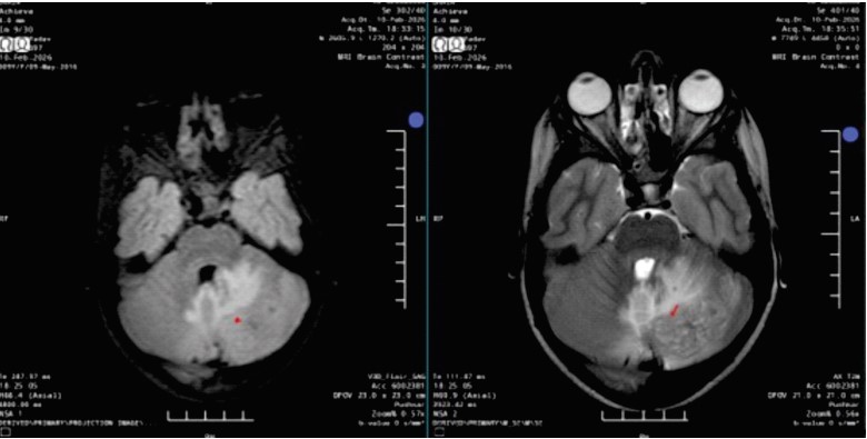
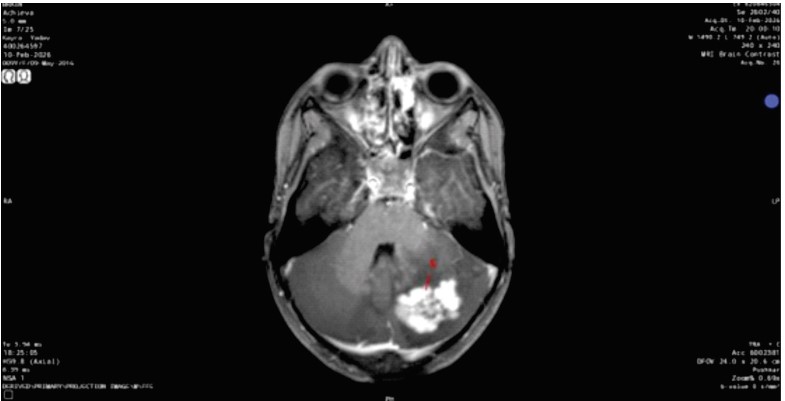
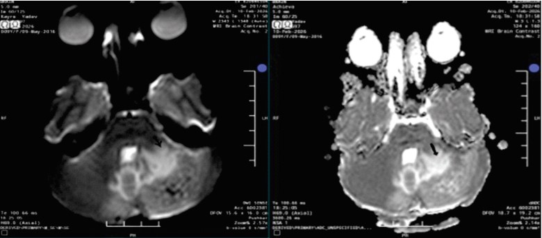
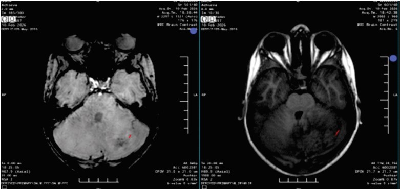
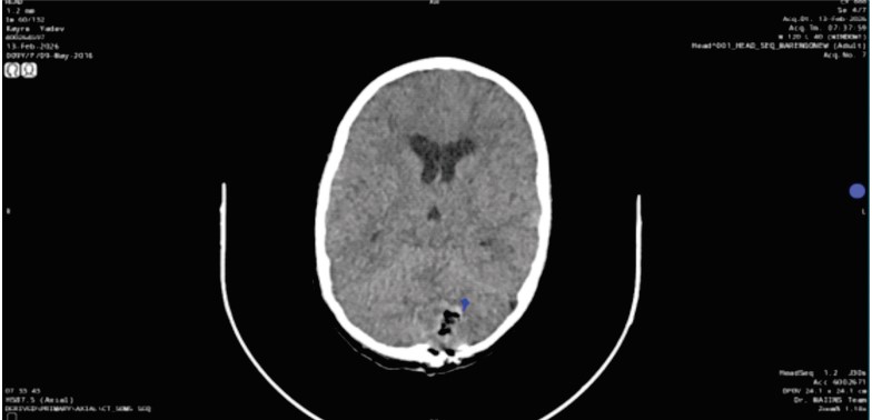
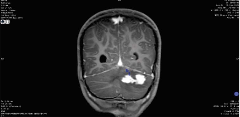
Introduction: Tuberculomas are most common lesions in India and can closely mimic neoplastic lesion, as ganglioglioma on imaging. Case Report: A 9-year-old female presented with severe headache in emergency department. Hence, she was referred to radiology department of MRI BRAIN and orbit imaging considering possibility of intracranial hypertension. On MRI, a solid-enhancing left cerebellar lesion was seen and it favored ganglioglioma. Patient was undertaken for the surgery, her histopathology suggested necrotizing granulomatous inflammation consistent with tuberculoma with no evidence of BIH. Conclusion: Cerebellar tuberculoma can present atypically and mimic neoplasm.
References
- 1. Rock RB, Olin M, Baker CA, Molitor TW, Peterson PK. Central nervous system tuberculosis: pathogenesis and clinical aspects. Clin Microbiol Rev. 2008;21(2):243-61.
- 2. Gupta RK, Jena A, Sharma A, Gujral RB, Husain N. MR imaging of intracranial tuberculomas. J Comput Assist Tomogr. 1988;12(2):280-5.
- 3. Bernaerts A, Vanhoenacker FM, Parizel PM, et al. Tuberculosis of the central nervous system: overview of neuroradiological findings. Eur Radiol. 2003;13(8):1876-90.
- 4. Louis DN, Perry A, Wesseling P, et al. The 2021 WHO Classification of Tumors of the CNS. Acta Neuropathol. 2021;142(4):567-85.
- 5. Lu ZR, Lai SW, Tsai YM. Unveiling a diagnostic challenge: Rare pulmonary tuberculosis and subsequent pleural tuberculoma mimicking relapse of Hodgkin lymphoma. Int J Infect Dis. 2025;161:108136. (5)
- 6. Khatri G D, Krishnan V, Antil N, Saigal G. Magnetic resonance imaging spectrum of intracranial tubercular lesions: one disease, many faces. Polish Journal of Radiology. 2018;83:628–639. doi:10.5114/pjr.2018.81408.
- 7. Chang KH, Han MH, Roh JK, et al. GdDTPA-enhanced MR imaging of intracranial tuberculosis. Radiology. 1990;176(2):563-7.
- 8. Koeller KK, Henry JM. Superficial gliomas: radiologic-pathologic correlation. Radiographics. 2001;21(6):1533-56.
- 9. Bhat AR, Wani MA, Kirmani AR. Histopathological Pattern and Outcome of Posterior Fossa Tumors in Children and Adults - A 20-Year Experience. Asian J Neurosurg. 2020 May 29;15(2):285-292. doi: 10.4103/ ajns.AJNS_120_19. PMID: 32656120; PMCID: PMC7335140.
Data Sharing Statement
There are no additional data available. All raw data and code are available upon request.
Funding
This research received no funding.
Author Contributions
All authors contributed significantly to the work and approve its publication.
Ethics Declaration
This article does not involve any human or animal subjects, and therefore does not require ethics approval.
Acknowledgements
We would like to express our gratitude to the patients, their families, and all those who have contributed to this study.
Conflicts of Interest
No conflicts of interest in this work.
About this article

Cite this article
Shallini Mittal, Rashmi Bhardwaj, Nikita Jajodia et. al, Cerebellar Tuberculoma mimicking Ganglioglioma in a pediatric patient: A Case reporting discordance of pathology-radiological imaging. Int. J Neuro Neurosurgery. 2026; 18(2): 68-72.
Licence:
Attribution-Non-commercial 4.0 International (CC BY-NC 4.0)This license enables reusers to distribute, remix, adapt, and build upon the material in any medium or format for noncommercial purposes only, and only so long as attribution is given to the creator.
| Received | Accepted | Published |
|---|---|---|
| May 25, 2026 | July 30, 2026 | August 30, 2026 |
DOI: https://doi.org/10.21088/ijnns.0975.0223.18226.4
Keywords
TuberculomaGangliogliomaPosterior Fossa TumorPediatric Brain TumorMRISolid EnhancementSearch for Similar Articles
Similar Articles
- Beyond Uremic Encephalopathy: Timely Thrombolysis in Artery of Percheron Infarct...
- Multiscale Imaging-Based Characterization of Human Cortical Organoids Generated...
- Outcomes of Emergency Department Based Ventilatory Management of Adult Patients...
- Muscle Weakness as the Genesis of Spinal Disease: Paradigm Shift in Understandin...
- Intramedullary Capillary Hemangioma of Conus Medullaris: Case Report and Review...
Article Level Metrics
Last UpdatedWednesday 28 October 2026, 14:27:51 (IST)
2680
Accesses
5
297
00
Citations
NA
NA
NA
Download citation
Article Keywords
Keyword Highlighting
Highlight selected keywords in the article text.
Timeline
| Received | May 25, 2026 |
| Accepted | July 30, 2026 |
| Published | August 30, 2026 |
licence
Attribution-Non-commercial 4.0 International (CC BY-NC 4.0)
This license enables reusers to distribute, remix, adapt, and build upon the material in any medium or format for noncommercial purposes only, and only so long as attribution is given to the creator.