Full Text (PDF)
International Journal of Neurology and Neurosurgery 18(2):p 57-67, May-August 2026. | DOI: https://doi.org/10.21088/ijnns.0975.0223.18226.3
Original Article
Multiscale Imaging-Based Characterization of Human Cortical Organoids Generated from Induced Pluripotent Stem Cells
Deepak Agrawal, Aparna Sharma, Sujata Mohanty, Prabhakar Singh
Author Information
Licence:
Attribution-Non-commercial 4.0 International (CC BY-NC 4.0)This license enables reusers to distribute, remix, adapt, and build upon the material in any medium or format for noncommercial purposes only, and only so long as attribution is given to the creator.
International Journal of Neurology and Neurosurgery 18(2):p 57-67, May-August 2026. | DOI: https://doi.org/10.21088/ijnns.0975.0223.18226.3
How Cite This Article:
Deepak Agrawal, Aparna Sharma, Sujata Mohanty et. al, Multiscale Imaging-Based Characterization of Human Cortical Organoids Generated from Induced Pluripotent Stem Cells. Int. J Neuro Neurosurgery. 2026; 18(2): 57-67.Timeline
Received : June 16, 2026
Accepted : August 02, 2026
Published : August 30, 2026
Abstract
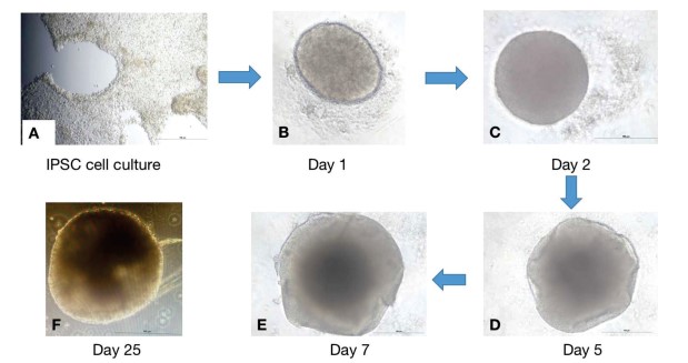
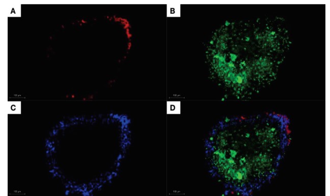
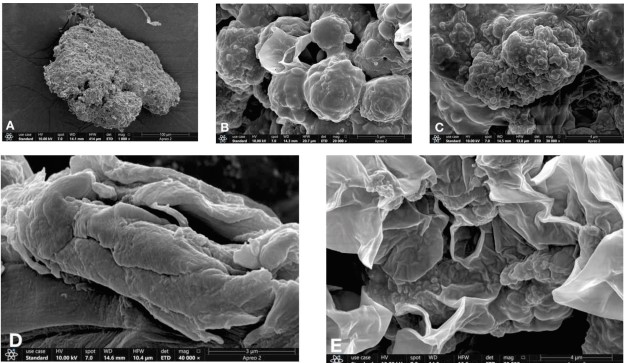
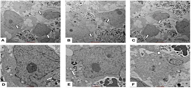
Human induced pluripotent stem cell (iPSC)-derived cortical organoids have emerged as transformative in vitro models for studying human neurodevelopment due to their remarkable ability to recapitulate early stages of corticogenesis in a three-dimensional environment. Unlike two-dimensional cultures, cortical organoids exhibit self-organizing properties that enable the formation of neuroepithelial regions, neuronal populations, and glial networks resembling the developing human cerebral cortex. In this study, we established a standardized and reproducible protocol for generating human cortical organoids from iPSCs and performed multiscale structural characterization using confocal microscopy, scanning electron microscopy (SEM), and transmission electron microscopy (TEM). Human iPSCs were cultured under feeder-free conditions and differentiated through embryoid body formation followed by neural induction and cortical differentiation. Mature organoids were analyzed to evaluate cellular organization, tissue morphology, and ultrastructural maturation. Confocal microscopy demonstrated well-organized cortical-like architecture characterized by uniform nuclear distribution, extensive GFAP-positive astroglial networks, and distinct neuronal cell bodies identified by NeuroTrace staining. SEM analysis revealed spherical organoids with dense and highly organized surface topology, neuroepithelial folding, and neurite-like projections suggestive of progressive neuronal connectivity and tissue remodeling. TEM confirmed neuronal maturation through the identification of well-defined neuronal cell bodies, intercellular junctions, and myelin sheath-like structure surrounding neuronal processes. These findings demonstrate that the generated cortical organoids successfully reproduce essential structural features of early human brain development. The integration of multimodal imaging techniques provides robust validation of organoid architecture and maturation, establishing this platform as a reliable and biologically relevant model for studying neurodevelopment and neurological disease mechanisms. This work further contributes toward the standardization of cortical organoid generation and characterization protocols for translational neuroscience research.
References
- 1. Alex AM, Buss C, Davis EP, Campos GL, Donald KA, Fair DA, Gaab N, Gao W, Gilmore JH, Girault JB, Grewen K, Groenewold NA, Hankin BL, Ipser J, Kapoor S, Kim P, Lin W, Luo S, Norton ES, O’Connor TG, Piven J, Qiu A, Rasmussen JM, Skeide MA, Stein DJ, Styner MA, Thompson PM, Wakschlag L, Knickmeyer R; ENIGMA ORIGINs Group (2023) Genetic influences on the developing young brain and risk for neuropsychiatric disorders. Biol Psychiatry 93(10):905–920. https://doi. org/10.1016/j.biopsych.2023.01.013
- 2. Cullen DK, Gordián-Vélez WJ, Struzyna LA, Jgamadze D, Lim J, Wofford KL, Browne KD, Chen HI (2019) Bundled three-dimensional human axon tracts derived from brain organoids. iScience 21:57–67. https://doi. org/10.1016/j.isci.2019.10.004
- 3. Di Lullo E, Kriegstein AR (2017) The use of brain organoids to investigate neural development and disease. Nat Rev Neurosci 18(10):573–584. https://doi.org/10.1038/nrn.2017.107
- 4. Di Stefano J, Di Marco F, Cicalini I, FitzGerald U, Pieragostino D, Verhoye M, Ponsaerts P, Van Breedam E (2025) Generation, interrogation, and future applications of microglia-containing brain organoids. Neural Regen Res 20(12):3448–3460.
- 5. Fan X, Hou K, Liu G, Shi R, Wang W, Liang G (2025) Strategies to overcome the limitations of current organoid technology—engineered organoids. J Tissue Eng 16:20417314251319475. https://doi.org/10.1177/20417314251319475
- 6. Giandomenico SL, Mierau SB, Gibbons GM, Wenger LMD, Masullo L, Sit T, Sutcliffe M, Boulanger J, Tripodi M, Derivery E, Paulsen O, Lakatos A, Lancaster MA (2019) Cerebral organoids at the air-liquid interface generate diverse nerve tracts with functional output. Nat Neurosci 22(4):669–679. https://doi. org/10.1038/s41593-019-0350-2
- 7. Kadoshima T, Sakaguchi H, Nakano T, Soen M, Ando S, Eiraku M, Sasai Y (2013) Selforganization of axial polarity, inside-out layer pattern, and species-specific progenitor dynamics in human ES cell-derived neocortex. Proc Natl Acad Sci USA 110(50):20284–20289. https://doi.org/10.1073/pnas.1315710110
- 8. Lancaster MA, Renner M, Martin CA, Wenzel D, Bicknell LS, Hurles ME, Homfray T, Penninger JM, Jackson AP, Knoblich JA (2013) Cerebral organoids model human brain development and microcephaly. Nature 501(7467):373–379. https://doi.org/10.1038/nature12517
- 9. Lee CT, Bendriem RM, Wu WW, Shen RF (2017) 3D brain organoids derived from pluripotent stem cells: promising experimental models for brain development and neurodegenerative disorders. J Biomed Sci 24:59. https://doi. org/10.1186/s12929-017-0362-8
- 10. Li ZX, Tu SX, Li YW, Hu ZB, Liu WT, Tao YC, Zhao L, Song NN, Chen JY, Zhang Q, Qi CC, Zhu HW, Ding YQ, Hu L (2026) Med13 is involved in the radial migration and contralateral projection of cortical neurons via PlxnA4. Commun Biol 9:394. https://doi. org/10.1038/s42003-026-09704-w
- 11. Pathak D, Sriram K (2023) Neuron-astrocyte omnidirectional signaling in neurological health and disease. Front Mol Neurosci 16:1169320. https://doi.org/10.3389/fnmol. 2023.1169320
- 12. Qian X, Nguyen HN, Song MM, Hadiono C, Ogden SC, Hammack C, Yao B, Hamersky GR, Jacob F, Zhong C, Yoon KJ, Jeang W, Lin L, Li Y, Thakor J, Berg DA, Zhang C, Kang E, Chickering M, Nauen D, Ho CY, Wen Z, Christian KM, Shi PY, Maher BJ, Wu H, Jin P, Tang H, Song H, Ming GL (2016) Brain-region-specific organoids using minibioreactors for modeling ZIKV exposure. Cell 165(5):1238–1254. https://doi.org/10.1016/j. cell.2016.04.032
- 13. Quadrato G, Nguyen T, Macosko EZ, Sherwood JL, Yang SM, Berger DR, Maria N, Scholvin J, Goldman M, Kinney JP, Boyden ES, Lichtman JW, Williams ZM, McCarroll SA, Arlotta P (2017) Cell diversity and network dynamics in photosensitive human brain organoids. Nature 545(7652):48–53. https://doi.org/10.1038/ nature22047
- 14. Rajagopalan V, Scott J, Habas PA, Kim K, Rousseau F, Glenn OA, Barkovich AJ, Studholme C (2011) Spatiotemporal morphometry of adjacent tissue layers with application to the study of sulcal formation. Med Image Comput Comput Assist Interv 14(Pt 2):476–483. https://doi.org/10.1007/978- 3-642-23629-7_58
- 15. Sultana of, Bandaru M, Islam MA, Reddy PH (2024) Unraveling the complexity of human brain: structure, function in healthy and disease states. Ageing Res Rev 100:102414. https://doi.org/10.1016/j.arr.2024.102414
- 16. Truong HD, Ge Z, Chng E, et al (2026) Strategies to control cellular spatial organization in microphysiological systems. Microsyst Nanoeng 12:85. https://doi.org/10.1038/ s41378-025-01141-9
- 17. Wang J, Qiao G, Tan H, Li MJ, Wang K, Xu J, Janowski M, Fu TM, Walczak P, Liang Y (2025) Intravital multimodal imaging of human cortical organoids for chronic stroke treatment in mice. bioRxiv. https://doi. org/10.1101/2025.07.05.663297
- 18. Xiang Y, Park IH (2024) Brain organoids: from unguided to regionalized to nucleusspecific. Life Med 3(2):lnae014. https://doi. org/10.1093/lifemedi/lnae014
- 19. Zhao X, Xu Z, Xiao L, Shi T, Xiao H, Wang Y, Li Y, Xue F, Zeng W (2021) Review on the vascularization of organoids and organoidson-a-chip. Front Bioeng Biotechnol 9:637048. https://doi.org/10.3389/fbioe.2021.637048
Data Sharing Statement
There are no additional data available. All raw data and code are available upon request.
Funding
This work was supported by the Indian Council of Medical Research (ICMR), Government of India.
Author Contributions
All authors contributed significantly to the work and approve its publication.
Ethics Declaration
This article does not involve any human or animal subjects, and therefore does not require ethics approval.
Acknowledgements
We would like to express our gratitude to the patients, their families, and all those who have contributed to this study.
Conflicts of Interest
No conflicts of interest in this work.
About this article

Cite this article
Deepak Agrawal, Aparna Sharma, Sujata Mohanty et. al, Multiscale Imaging-Based Characterization of Human Cortical Organoids Generated from Induced Pluripotent Stem Cells. Int. J Neuro Neurosurgery. 2026; 18(2): 57-67.
Licence:
Attribution-Non-commercial 4.0 International (CC BY-NC 4.0)This license enables reusers to distribute, remix, adapt, and build upon the material in any medium or format for noncommercial purposes only, and only so long as attribution is given to the creator.
| Received | Accepted | Published |
|---|---|---|
| June 16, 2026 | August 02, 2026 | August 30, 2026 |
DOI: https://doi.org/10.21088/ijnns.0975.0223.18226.3
Keywords
Human induced pluripotent stem cells (hiPSCs)Cortical organoidsBrain organoidsNeurodevelopmentStructural characterizationSearch for Similar Articles
Similar Articles
- Beyond Uremic Encephalopathy: Timely Thrombolysis in Artery of Percheron Infarct...
- Cerebellar Tuberculoma Mimicking Ganglioglioma in a Pediatric Patient: A Case Re...
- Outcomes of Emergency Department Based Ventilatory Management of Adult Patients...
- Muscle Weakness as the Genesis of Spinal Disease: Paradigm Shift in Understandin...
- Intramedullary Capillary Hemangioma of Conus Medullaris: Case Report and Review...
Article Level Metrics
Last UpdatedWednesday 28 October 2026, 14:30:19 (IST)
2680
Accesses
5
297
00
Citations
NA
NA
NA
Download citation
Article Keywords
Keyword Highlighting
Highlight selected keywords in the article text.
Timeline
| Received | June 16, 2026 |
| Accepted | August 02, 2026 |
| Published | August 30, 2026 |
licence
Attribution-Non-commercial 4.0 International (CC BY-NC 4.0)
This license enables reusers to distribute, remix, adapt, and build upon the material in any medium or format for noncommercial purposes only, and only so long as attribution is given to the creator.