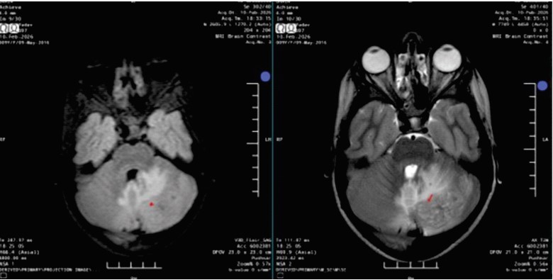
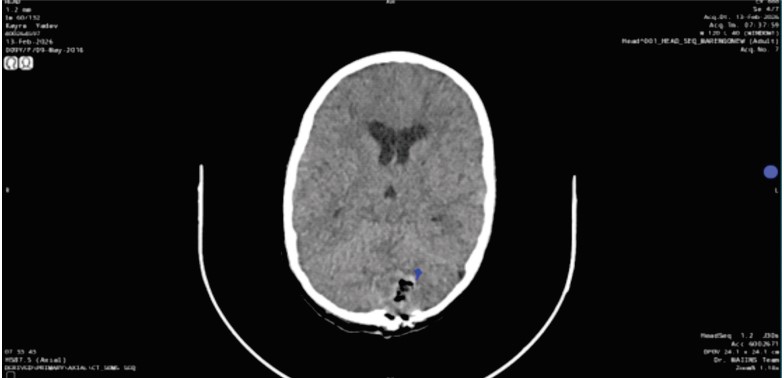
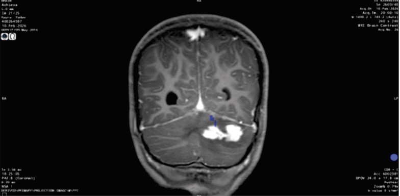
Description: Axial T2/FLAIR image shows a heterogeneously hyperintense lesion in the left cerebellar hemisphere with surrounding edema
Heading
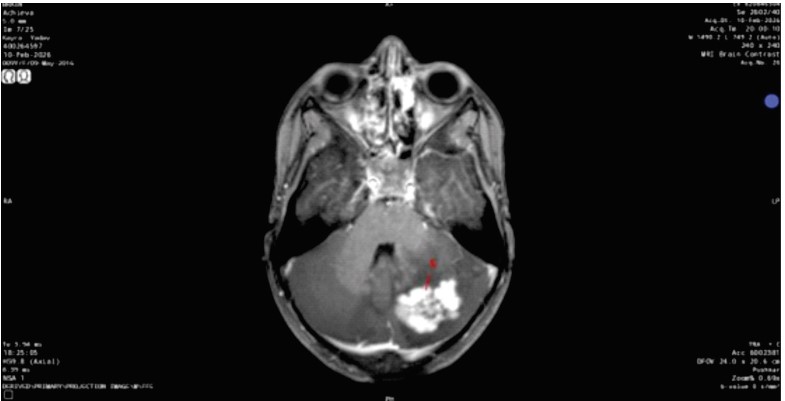
Description: Axial post-contrast T1-weighted image demonstrates predominantly solid avid enhancement of the left cerebellar lesion
(marked in red as c)
Heading
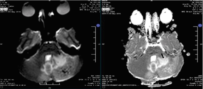
Description: DWI/ADC maps show no diffusion restriction within the lesion (DW arrow, ADC thick solid arrow)
Heading
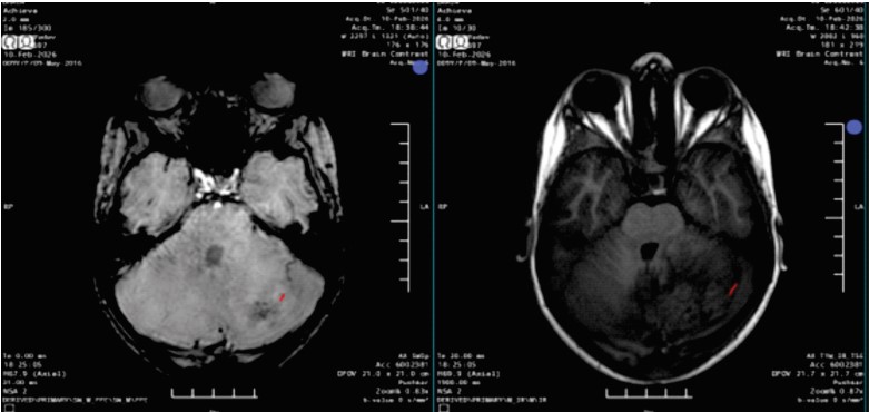
Description: SWI demonstrates susceptibility blooming foci within the lesion (Red mark)
Heading
Description: Non-contrast CT shows post-operative changes in posterior fossa (hemorrhage and air loculi — marked blue)
Heading
Description: Coronal post-contrast T1-weighted image demonstrates solid enhancing lesion in left cerebellum (Marked e)