Full Text (PDF)
Journal of Dermatology 8(2):p 107-111, July-December 2023. | DOI: NA
Case Report
Case of Lichen Planus Pigmentosus with Atypical Presentation: A Case Report
Akshay Arvind Patankar, Akshata Mane, Manjaree Morgaonkar
Author Information
Licence:
Attribution-Non-commercial 4.0 International (CC BY-NC 4.0)This license enables reusers to distribute, remix, adapt, and build upon the material in any medium or format for noncommercial purposes only, and only so long as attribution is given to the creator
Journal of Dermatology 8(2):p 107-111, July-December 2023. | DOI: NA
How Cite This Article:
Patankar AA, Mane A, Morgaonkar M. Case of Lichen Planus Pigmentosus with Atypical Presentation: A Case Report. RFP J Dermatol. 2023;8(2):107–111.Timeline
Received : May 05, 2023
Accepted : June 15, 2023
Published : December 30, 2023
Abstract
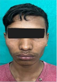
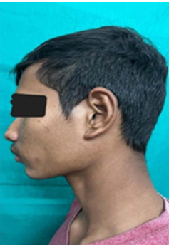
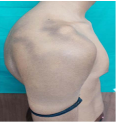
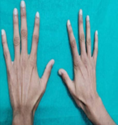
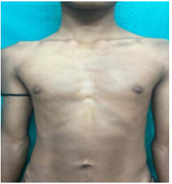
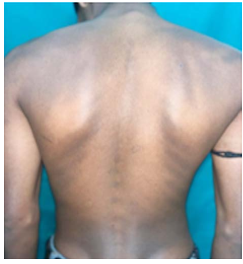
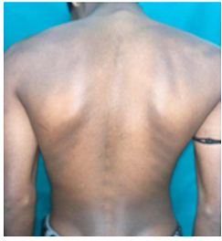
Lichen planus pigmentosus (LPP) is a chronic pigmentary disorder that shows diffuse or reticulated hyperpigmented, dark brown macules and patches on the sun-exposed areas such as the face, neck and other flexural folds. Clinically, it is different from classical lichen planus because LPP has a longer clinical course and it manifests with dark brown macules. In case of LPP, involvement of the scalp, nail or mucosal area is rare. The histopathological findings of the lesions show an atrophic epidermis, the presence of melanophages and a vacuolar alteration of the basal cell layer with a sparse lymphohistiocytic lichenoid infiltration. Here we report a case of lichen planus pigmentosus over sun protected areas
References
- 2. Ghosh A, Coondoo A. Lichen planus pigmentosus: The controversial consensus. Indian journal of dermatology. 2016 Sep;61(5):482.
- 3. Kanwar AJ, Dogra S, Handa S, Parsad D, Radotra BD. A study of 124 Indian patients with lichen planus pigmentosus. Clin Exp Dermatol. 2003;28:481–5. [PubMed: 12950331]
- 4. Rieder E, Kaplan J, Kamino H, Sanchez M, Pomeranz MK. Lichen planus pigmentosus. Dermatol Online J. 2013;19:20713. [PubMed: 24365004]
- 5. Vachiramon V, Suchonwanit P, Thadanipon K. Bilateral linear lichen planus pigmentosus associated with hepatitis C virus infection. Case Rep Dermatol. 2010;2:169–172. [PMCID: PMC2974975] [PubMed: 21060775]
- 6. Gupta D, Thappa DM. Dermatoses due to Indian cultural practices. Indian J Dermatol. 2015;60:3–12. [PMCID: PMC4318059] [PubMed: 25657390]
- 7. Al-Mutairi N, El-Khalawany M. Clinicopathological characteristics of lichen planus pigmentosus and its response to tacrolimus ointment: an open label, nonrandomized, prospective study. J Eur Acad Dermatol Venereol 2010; 24: 535–540.
- 8. Kumar YH, Babu AR. Segmental lichen planus pigmentosus: An unusual presentation. Indian Dermatol Online J. 2014;5:157–9. [PMCID: PMC4030343] [PubMed: 24860750]
- 9. Cho S, Whang KK. Lichen planus pigmentosus presenting in zosteriform pattern. J Dermatol. 1997;24:193–7. [PubMed: 9114619]
- 10. Hong S, Shin JH, Kang HY. Two cases of lichen planus pigmentosus presenting with a linear pattern. J Korean Med Sci. 2004;19:152–4. [PMCID: PMC2822256] [PubMed: 14966361]
- 11. Vineet R, Sumit S, Garg VK, Nita K. Lichen planus pigmentosusin linear and zosteriform pattern along the lines of Blaschko. Dermatol Online J. 2015;21 pii: 13030/qt4rk2w3rm. [PubMed: 26632802]
- 12. Pock L, Jelınkova L, Drlık L, et al. Lichen planus pigmentosus-inversus. J Eur Acad Dermatol Venereol 2001; 15: 452–454.
- 13. Robles Méndez JC, Rizo Frías P, Herz Ruelas ME, Pandya AG, OcampoCandiani J. Lichen planus pigmentosus and its variants: review and update. International journal of dermatology. 2018 May;57(5):505-14.
- 14. Murzaku EC, Bronsnick T, Rao BK. Axillary lichen planus pigmentosus-inversus: dermoscopic clues of a rare entity. Diagnosis: lichen planus pigmentosus (LPP). J. Am. Acad. Dermatol. 2014; 71: e119–20. \
- 15. Sonthalia S, Errichetti E, Kaliyadan F, Jha AK, Lallas A. Dermoscopy of lichen planus pigmentosus in Indian patients–Pitfalls to avoid. Indian Journal of Dermatology, Venereology and Leprology. 2018 May 1;84:311.
- 16. Kumarasinghe SP, Pandya A, Chandran V, Rodrigues M, Dlova NC, Kang HY, Ramam M, Dayrit JF, Goh BK, Parsad D. A global consensus statement on ashy dermatosis, erythema dyschromicumperstans, lichen planus pigmentosus, idiopathic eruptive macular pigmentation, and Riehl's melanosis. International Journal of Dermatology. 2019 Mar;58(3):263-72.
- 17. Robles Méndez JC, Rizo Frías P, Herz Ruelas ME, Pandya AG, OcampoCandiani J. Lichen planus pigmentosus and its variants: review and update. International journal of dermatology. 2018 May;57(5):505-14.
Data Sharing Statement
There are no additional data available
Funding
This research received no funding
Author Contributions
All authors contributed significantly to the work and approve its publication
Ethics Declaration
This article does not involve any human or animal subjects, and therefore does not require ethics approval
Acknowledgements
Information Not Provided
Conflicts of Interest
No conflicts of interest in this work.
About this article

Cite this article
Patankar AA, Mane A, Morgaonkar M. Case of Lichen Planus Pigmentosus with Atypical Presentation: A Case Report. RFP J Dermatol. 2023;8(2):107–111.
Licence:
Attribution-Non-commercial 4.0 International (CC BY-NC 4.0)This license enables reusers to distribute, remix, adapt, and build upon the material in any medium or format for noncommercial purposes only, and only so long as attribution is given to the creator
| Received | Accepted | Published |
|---|---|---|
| May 05, 2023 | June 15, 2023 | December 30, 2023 |
DOI: NA
Keywords
Lichen Planus PigmentosusPigment IncontinenceSun protected areasSearch for Similar Articles
Similar Articles
- Role of Topical Cholecalciferol Granules in the Recovery and Management of Therm...
- Chitosan Liquid Wound Dressing in the Use of Deep Scald Burns
- Role of Biocellulose Occlusive Dressing in Partial Thickness Burn Wound Manageme...
- Artificial Intelligence in the Digital Transformation of Dermatology: Current E...
- Artificial Intelligence in Dermatology in India: A PRISMA-Compliant Systematic...
Article Level Metrics
Last UpdatedSaturday 05 September 2026, 18:20:59 (IST)
1128
Accesses
10
109
00
Citations
NA
NA
NA
Download citation
Article Keywords
Keyword Highlighting
Highlight selected keywords in the article text.
Timeline
| Received | May 05, 2023 |
| Accepted | June 15, 2023 |
| Published | December 30, 2023 |
licence
Attribution-Non-commercial 4.0 International (CC BY-NC 4.0)
This license enables reusers to distribute, remix, adapt, and build upon the material in any medium or format for noncommercial purposes only, and only so long as attribution is given to the creator