Full Text (PDF)
Journal of Pharmaceutical and Medicinal Chemistry 9(1):p 33-35, January-June 2023. | DOI: https://doi.org/10.21088/jpmc.2395.6615.9123.3
Case Report
Bullous Pemphigoid as an Adverse Reaction to PD-1 Inhibitor Avelumab: A Case Report
Divya Yadav, Manisha Nijhawan, Sankalp Awasthi, Akshay Goyal
Author Information
Licence:
Attribution-Non-commercial 4.0 International (CC BY-NC 4.0)This license enables reusers to distribute, remix, adapt, and build upon the material in any medium or format for noncommercial purposes only, and only so long as attribution is given to the creator.
Journal of Pharmaceutical and Medicinal Chemistry 9(1):p 33-35, January-June 2023. | DOI: https://doi.org/10.21088/jpmc.2395.6615.9123.3
How Cite This Article:
Divya Yadav, Manisha Nijhawan, Sankalp Awasthi, et al./Bullous Pemphigoid as an Adverse Reaction to PD-1 inhibitor Avelumab: A Case Report/J Pharmaceut Med Chem. 2023;9(1): 33-35.Timeline
Received : April 17, 2023
Accepted : May 25, 2023
Published : June 25, 2023
Abstract
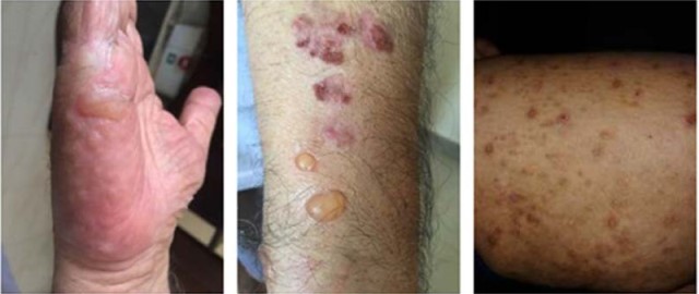
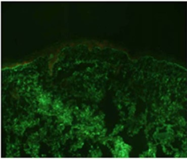
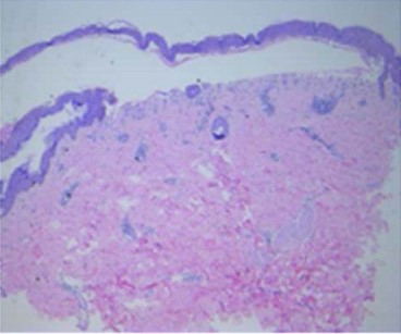
Checkpoint inhibitors targeting CTLA4 (Cytotoxic T-lymphocyte antigen-4), PD-1 (programmed death)1 on lymphocytes and PD-L1 (programmed death ligand)1 on tumors cells are novel and promising treatment options for different types of cancer. But these agents are double edged swords, they undoubtedly robust the anti-tumor response but also throttle up the normal immunologic homeostasis. Autoimmune adverse reactions are very common with checkpoint inhibitors. We present a case of bullous pemphigoid, as an adverse reactions to Avelumab treated urinary bladder cancer with liver metastasis.
References
- 1. Belum VR, Benhuri B, Postow MA, et al. Characterisation and management of dermatologic adverse events to agents targeting the PD-1 receptor. Eur J Cancer. 2016;60(2016):12–25. doi: 10.1016/j. ejca.2016.02.010.
- 2. de Golian E, Kwong BY, Swetter SM, Pugliese SB. Cutaneous Complications of Targeted Melanoma Therapy. Curr Treat Options Oncol. 2016;17(11). doi:10.1007/s11864-016-0434-0.
- 3. Friedman CF, Proverbs-Singh TA, and Postow MA, Treatment of the Immune-Related Adverse Effects of Immune Checkpoint Inhibitors: A Review. JAMA Oncol, 2016 2(10): p. 1346–1353.
- 4. Kershenovich R, Hodak E, Mimouni D. Diagnosis and classification of pemphigus and bullous pemphigoid. Autoimmun Rev. 2014;13(4–5):477– 481. doi:10.1016/j.autrev.2014.01.011.
- 5. Burton JL, Harman RR, Peachey RD, Warin RP et al. Azathioprine plus prednisone in treatment of pemphigoid. Br Med J. 1978 10 28;2(6146):1190–1.
- 6. Eskin-Schwartz M, David M, Mimouni D et al. Mycophenolatemofetil for the management of autoimmune bullous diseases. DermatolClin. 2011 Oct;29(4):555–9.
Data Sharing Statement
There are no additional data available. All raw data and code are available upon request.
Funding
This research received no funding.
Author Contributions
All authors contributed significantly to the work and approve its publication.
Ethics Declaration
This article does not involve any human or animal subjects, and therefore does not require ethics approval.
Acknowledgements
We would like to express our gratitude to the patients, their families, and all those who have contributed to this study.
Conflicts of Interest
No conflicts of interest in this work.
About this article

Cite this article
Divya Yadav, Manisha Nijhawan, Sankalp Awasthi, et al./Bullous Pemphigoid as an Adverse Reaction to PD-1 inhibitor Avelumab: A Case Report/J Pharmaceut Med Chem. 2023;9(1): 33-35.
Licence:
Attribution-Non-commercial 4.0 International (CC BY-NC 4.0)This license enables reusers to distribute, remix, adapt, and build upon the material in any medium or format for noncommercial purposes only, and only so long as attribution is given to the creator.
| Received | Accepted | Published |
|---|---|---|
| April 17, 2023 | May 25, 2023 | June 25, 2023 |
DOI: https://doi.org/10.21088/jpmc.2395.6615.9123.3
Keywords
Bullous pemphigoidAvelumabBladder carcinomaSearch for Similar Articles
Similar Articles
- Pharmaceutical Care in Primary Healthcare: A Study of Nurses
- Artificial Intelligence in Pharmaceutical Research and Development: From Target...
- Developing a Structured Teaching Program for Precision Medicine in Nursing Pract...
- Comprehensive Scientific Review on the Natural Process of Aging, Anti-Aging Mech...
- Formulation and Evaluation of Curcumin Lozenges to Treat Throat Infections
Article Level Metrics
Last UpdatedMonday 26 October 2026, 22:25:38 (IST)
1383
Accesses
5
118
00
Citations
NA
NA
NA
Download citation
Article Keywords
Keyword Highlighting
Highlight selected keywords in the article text.
Timeline
| Received | April 17, 2023 |
| Accepted | May 25, 2023 |
| Published | June 25, 2023 |
licence
Attribution-Non-commercial 4.0 International (CC BY-NC 4.0)
This license enables reusers to distribute, remix, adapt, and build upon the material in any medium or format for noncommercial purposes only, and only so long as attribution is given to the creator.