Full Text (PDF)
Indian Journal of Research in Anthropology 11(1):p 43-53, Jan-June 2025. | DOI: https://doi.org/10.21088/ijra.2454.9118.11125.5
Original Article
Anthropometric Estimation of Metacarpophalangeal Profile in Individuals with Brachydactyly: A Radiometric Study
Inusha Panigrahi, Shalini Dhiman, Harvinder Kaur, Suresh Kumar Sharma, Shifali Gupta
Author Information
Licence:
Attribution-Non-commercial 4.0 International (CC BY-NC 4.0)This license enables reusers to distribute, remix, adapt, and build upon the material in any medium or format for noncommercial purposes only, and only so long as attribution is given to the creator.
Indian Journal of Research in Anthropology 11(1):p 43-53, Jan-June 2025. | DOI: https://doi.org/10.21088/ijra.2454.9118.11125.5
How Cite This Article:
Shalini Dhiman, Inusha Panigrahi, Harvinder Kaur et al. Anthropometric Estimation of Metacarpophalangeal Profile in Individuals with Brachydactyly: A Radiometric Study. Ind J Res Anthropol 2025; 11(1): 43-53.Timeline
Received : January 01, 2025
Accepted : March 06, 2025
Published : June 30, 2025
Abstract
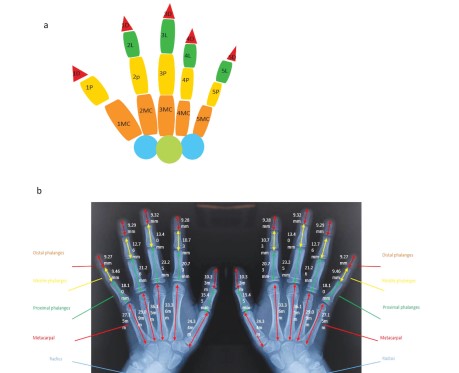
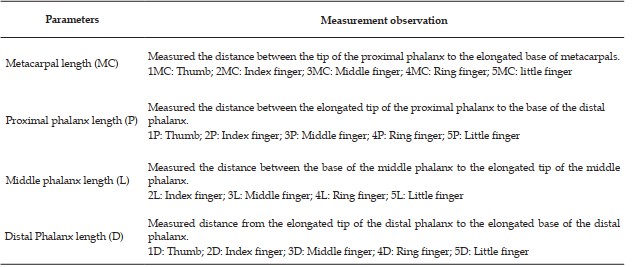
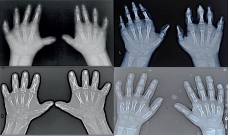
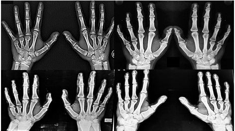
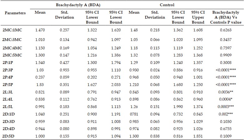
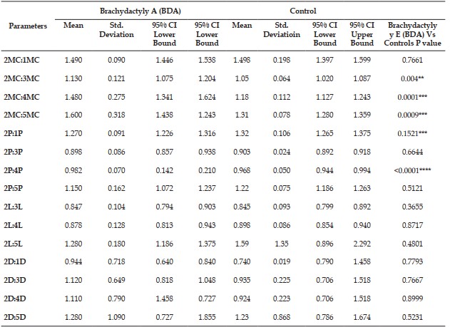
Background: The bone length of paediatric patients varies with respect to normal individuals. Patients were mainly affected with congenital autopod anomalies such as brachydactyly, syndactyly, and polydactyly. Brachydactyly with short bones are further divided into different types: Brachydactyly A, brachydactyly B, brachydactyly C, brachydactyly D, and brachydactyly E. Aim: This study aims to classify the types of brachydactyly by measuring metacarpophalangeal profile patients: A radiometric study Objectives: Classify the types of brachydactyly patients and compare them with the controls based on X-rays of hands. Material: A cross-sectional study was conducted on the children at a tertiary care center from July 2020 to March 2024. Metacarpal and phalangeal lengths of little, ring, middle, index, and thumb fingers on X-ray films of patients and controls were measured by digimatic calipers. The recorded data were analysed by SPSS version 26 and Graph Pad Prisma 8.2. Result: We found highly significant p values in ratios of phalangeal lengths 2P:3P, 2P:4P, 2P:5P, 2L:3L, 2L:4L, 2L: 5L ( P; Proximal phalanx length , L: middle phalanx length). This predicted abnormal or short/missing proximal, middle, and distal phalanges of individuals with brachydactyly type A. We found highly significant p values in ratios of (MC: Metacarpal length) 2MC: 3MC, 2MC:4MC, 2MC:5MC, and 2P:4P in brachydactyly type E. Conclusion: This predicted short 3rd, 4th, 5th metacarpals and 4th proximal phalanges in comparison to controls. Thus, X-ray analysis is the gold standard tool for evaluating the different types of brachydactyly. The research emphasizes the importance of hand anthropometry in understanding anatomical changes and the diversity of brachydactyly. Key Message: This study helps to classify the rare genetic disease in pediatric patients and better understanding for the genetic counselling. Also helpful in the surgical treatment of hands in complex cases.
References
- 1. Jurcă M.C., Bembea M.A., Şandor M.I., Zaha D.C., Negrean R.A., Vesa C.M., Jurcă A.A., Moisa F.C., Vicaş L.G., Paul C., Cheregi S.D. Congenital anomalies of digits—A clinicalepidemiological study of 301 patients. Rom J Morphol Embryol. 2019 Jan 1; 60(4): 1221-6.
- 2. Hall C.M. International nosology and classification of constitutional disorders of bone (2001). American journal of medical genetics. 2002 Nov 15; 113(1): 65-77. https://doi.org/10.1002/ajmg.10828
- 3. Unger S., Ferreira C.R., Mortier G.R., et al. Nosology of genetic skeletal disorders: 2023 revision. American Journal of Medical Genetics Part A. 2023 May; 191(5): 1164-209. https:// doi.org/10.1002/ajmg.a.63132
- 4. Pereda A., Garin I., Garcia-Barcina M., et al. Brachydactyly E: isolated or as a feature of a syndrome. Orphanet journal of rare diseases. 2013 Dec; 8: 1-4. https://doi.org/10.1186/1750- 1172-8-141
- 5. Temtamy S.A., Aglan M.S. Brachydactyly. Orphanet journal of rare diseases. 2008 Dec;3:1-6. https://doi.org/10.1186/1750-1172-3-15
- 6. David A., Vincent M., Quéré M.P., Lefrançois T., Frampas E. Isolated and syndromic brachydactylies: Diagnostic value of hand X-rays. Diagnostic and Interventional Imaging. 2015 May 1; 96(5): 443-8. https://doi.org/10.1016/j.diii.2014.12.007
- 7. Leung A.O., Poon A.C., Wang X., et al. Suppression of apoptosis impairs phalangeal joint formation in the pathogenesis of brachydactyly type A1. Nature Communications. 2024 Mar 12;15(1):2229. https://doi.org/10.1038/s41467-024-45053-0
- 8. Sun J., Yang N., Xu Z., Cheng H., Zhang X. A novel heterozygous mutation in PTHLH causing autosomal dominant brachydactyly type E complicated with short stature. Molecular Genetics & Genomic Medicine. 2024 Feb; 12(2): e2393.https://doi.org/10.1002/mgg3.2393
- 9. Zaib T, Rashid H, Khan H, Zhou X, Sun P. Recent advances in syndactyly: Basis, current status and future perspectives. Genes. 2022 Apr 27; 13(5): 771. https://doi.org/10.3390/genes13050771
- 10. Ahmad Z, Liaqat R, Palander O, et al. Genetic overview of postaxial polydactyly: updated classification. Clinical Genetics. 2023 Jan; 103(1): 3-15. https://doi.org/10.1111/cge.14224
- 11. Manning J.T., Trivers I.V. R.L., Thornhill R., Singh D. The 2nd: 4th digit ratio and asymmetry of hand performance in Jamaican children. Laterality: Asymmetries of Body, Brain and Cognition. 2000 Apr 1; 5(2): 121-32. https://doi.org/10.1080/00222937100770081
- 12. Sephien A., Bethel C.F., Doyle C.M., Gulick D., Smith C.J., Schwartz-Fernandes FA. Morphometric analysis of the second through fifth metacarpal through posteroanterior X-Rays. Clinical Anatomy. 2020 Oct; 33(7):1014-8. https://doi.org/10.1002/ca.23528
- 13. Foley D.P., Cox C.T., Foley A.S., et al. Predicting metacarpal length using paired ratios with bilateral X-ray films. SAGE Open Medicine. 2021 Dec; 9 https://doi.org/10.1177/20503121211064177
Data Sharing Statement
There are no additional data available. All raw data and code are available upon request.
Funding
This research received no funding.
Author Contributions
All authors contributed significantly to the work and approve its publication.
Ethics Declaration
This article does not involve any human or animal subjects, and therefore does not require ethics approval.
Acknowledgements
We would like to express our gratitude to the patients, their families, and all those who have contributed to this study.
Conflicts of Interest
No conflicts of interest in this work.
About this article

Cite this article
Shalini Dhiman, Inusha Panigrahi, Harvinder Kaur et al. Anthropometric Estimation of Metacarpophalangeal Profile in Individuals with Brachydactyly: A Radiometric Study. Ind J Res Anthropol 2025; 11(1): 43-53.
Licence:
Attribution-Non-commercial 4.0 International (CC BY-NC 4.0)This license enables reusers to distribute, remix, adapt, and build upon the material in any medium or format for noncommercial purposes only, and only so long as attribution is given to the creator.
| Received | Accepted | Published |
|---|---|---|
| January 01, 2025 | March 06, 2025 | June 30, 2025 |
DOI: https://doi.org/10.21088/ijra.2454.9118.11125.5
Keywords
Acral shorteningBrachymetacarpyDigimatic CaliperFingersHand AnomaliesHuman diversityMetacarpalsPhalangesSearch for Similar Articles
Similar Articles
- Contemporary Issues and Challenges in Women’s Weavers in the Varanasi Silk Saree...
- Understanding through Traversing the Corridors of History: Richness of Indian He...
- Challenges and Opportunities in Digital Preservation of Intangible Culture Herit...
- Assessment of Nutritional Indicators: A Comparative Study among Pre-School Child...
- Prenatal and Postnatal Health-Seeking Behaviour among the Muria Tribe of Bastar...
Article Level Metrics
Last UpdatedTuesday 08 September 2026, 20:57:39 (IST)
1819
Accesses
12
413
00
Citations
NA
NA
NA
Download citation
Article Keywords
Keyword Highlighting
Highlight selected keywords in the article text.
Timeline
| Received | January 01, 2025 |
| Accepted | March 06, 2025 |
| Published | June 30, 2025 |
licence
Attribution-Non-commercial 4.0 International (CC BY-NC 4.0)
This license enables reusers to distribute, remix, adapt, and build upon the material in any medium or format for noncommercial purposes only, and only so long as attribution is given to the creator.