Full Text (PDF)
Journal of Dermatology 10(2):p 47-49, July-December 2025. | DOI: https://doi.org/10.21088/jd.2582.3582 10225.1
Editorial
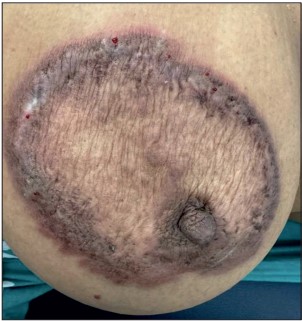
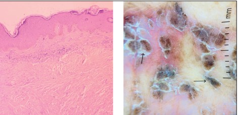
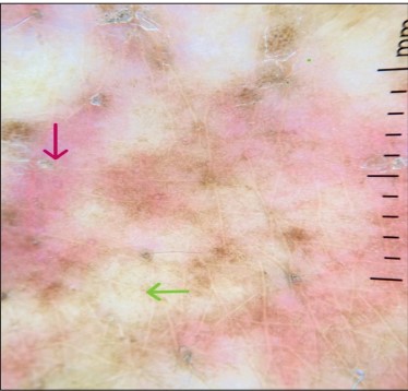
Annular Keloid Mimicking Granuloma Annulare: A Diagnostic Challenge
Pragya A. Nair, Priya Ghoghara, Shree Dhanani
Author Information
Licence:
Attribution-Non-commercial 4.0 International (CC BY-NC 4.0)This license enables reusers to distribute, remix, adapt, and build upon the material in any medium or format for noncommercial purposes only, and only so long as attribution is given to the creator.
Journal of Dermatology 10(2):p 47-49, July-December 2025. | DOI: https://doi.org/10.21088/jd.2582.3582 10225.1
How Cite This Article:
Priya Ghoghara, Shree Dhanani, Pragya A. Nair. Annular Keloid Mimicking Granuloma Annulare: A Diagnostic Challenge. RFP Jr of Drea 2025; 10(2): 47-49.Timeline
Received : September 16, 2025
Accepted : November 18, 2025
Published : December 25, 2025
Abstract
NA
References
- 1. Hunasgi S, Koneru A, Vanishree M, Shamala R. Keloid: A case report and review of pathophysiology and differences between keloid and hypertrophic scars. Journal of Oral and Maxillofacial Pathology: JOMFP. 2013 ;17(1):116.
- 2. Nangole FW, Agak GW. Keloid pathophysiology: fibroblast or inflammatory disorders?. JPRAS open. 2019;22:44-54.
- 3. Schmieder SJ, Harper CD, Schmieder GJ. Granuloma Annulare. [Updated 2023 Jun 21]. In: StatPearls [Internet].
- 4. Yoo MG, Kim IH. Keloids and hypertrophic scars: characteristic vascular structures visualized by using dermoscopy. Annals of dermatology. 2014 ;26(5):603-9.
- 5. Angit C, Clayton TH. Lightning-strike granuloma. Clin Exp Dermatol. 2009;34(7):e485-6.
Data Sharing Statement
There are no additional data available. All raw data and code are available upon request.
Funding
This research received no funding.
Author Contributions
All authors contributed significantly to the work and approve its publication.
Ethics Declaration
This article does not involve any human or animal subjects, and therefore does not require ethics approval.
Acknowledgements
We would like to express our gratitude to the patients, their families, and all those who have contributed to this study.
Conflicts of Interest
No conflicts of interest in this work.
About this article

Cite this article
Priya Ghoghara, Shree Dhanani, Pragya A. Nair. Annular Keloid Mimicking Granuloma Annulare: A Diagnostic Challenge. RFP Jr of Drea 2025; 10(2): 47-49.
Licence:
Attribution-Non-commercial 4.0 International (CC BY-NC 4.0)This license enables reusers to distribute, remix, adapt, and build upon the material in any medium or format for noncommercial purposes only, and only so long as attribution is given to the creator.
| Received | Accepted | Published |
|---|---|---|
| September 16, 2025 | November 18, 2025 | December 25, 2025 |
DOI: https://doi.org/10.21088/jd.2582.3582 10225.1
Keywords
Search for Similar Articles
Similar Articles
- Delving into Wolf’s Isotopic Phenomenon: Tale of Two Cases of Psoriasis Localize...
- Pachydermoperiostosis, Complete Form: A Case Report of Rare Occurrence
- Role of Cyclical Negative Pressure Wound Therapy in Thermal Burns
- Behcets Disease in an Immunocompromised Female
- Tale of an Atypical Herpes Zoster Mimicking Ecthyma and Perforating Dermatosis i...
Article Level Metrics
Last UpdatedThursday 01 October 2026, 05:35:14 (IST)
1528
Accesses
24
192
00
Citations
NA
NA
NA
Download citation
Article Keywords
Keyword Highlighting
Highlight selected keywords in the article text.
Timeline
| Received | September 16, 2025 |
| Accepted | November 18, 2025 |
| Published | December 25, 2025 |
licence
Attribution-Non-commercial 4.0 International (CC BY-NC 4.0)
This license enables reusers to distribute, remix, adapt, and build upon the material in any medium or format for noncommercial purposes only, and only so long as attribution is given to the creator.