Full Text (PDF)
Journal of Dermatology 10(2):p 65-70, July-December 2025. | DOI: https://doi.org/10.21088/jd.2582.3582 10225.4
Case Report
Delving into Wolf’s Isotopic Phenomenon: Tale of Two Cases of Psoriasis Localized to the site of Scars
Vijay Deepak Joshi, Ujjwal Sanjay kumar Rathi, Vidya Kharkar, Ketki Bhoite
Author Information
Licence:
Attribution-Non-commercial 4.0 International (CC BY-NC 4.0)This license enables reusers to distribute, remix, adapt, and build upon the material in any medium or format for noncommercial purposes only, and only so long as attribution is given to the creator.
Journal of Dermatology 10(2):p 65-70, July-December 2025. | DOI: https://doi.org/10.21088/jd.2582.3582 10225.4
How Cite This Article:
Ujjwal Sanjay kumar Rathi, Vidya Kharkar, Vijay Joshi et al. Delving into Wolf’s Isotopic Phenomenon: Tale of Two Cases of Psoriasis Localized to the site of Scars. RFP Jr of Drea 2025; 10(2): 65-70.Timeline
Received : May 21, 2025
Accepted : July 21, 2025
Published : December 25, 2025
Abstract
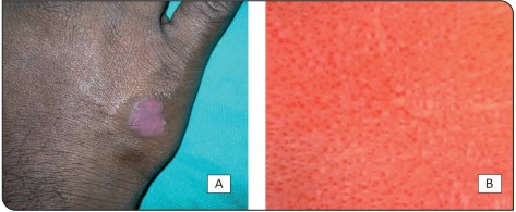
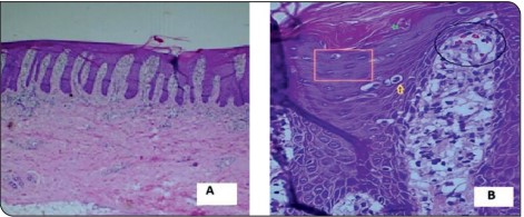
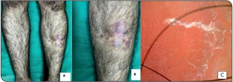
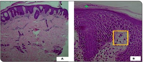
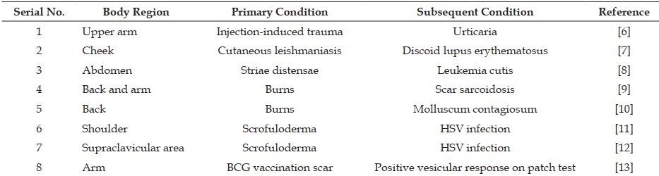
Wolf’s isotopic response refers to the occurrence of a new dermatosis at the site of a previously healed and unrelated skin condition. While commonly associated with viral triggers such as herpes simplex or zoster, non-viral initiators are rarely reported. We present two unique cases of psoriasis developing exclusively at sites of old scars from non-viral causes. The first case involved a 33-year-old male who developed a psoriatic plaque over a thermal burn scar on the dorsum of the hand. The second case featured a 28-year-old male with psoriatic lesions localized to an atrophic scar following a traumatic injury to the shin. Both cases lacked involvement of other body areas. Diagnosis was confirmed by clinical features, dermoscopy, and histopathology. These cases highlight an atypical presentation of Wolf’s isotopic phenomenon and underscore the potential role of localized immune dysregulation and scar-mediated vulnerability in the development of secondary dermatoses.
References
- 1. Mahajan R, De D, Saikia UN. Wolf’s Isotopic Response: Report of a Case and Review of Literature. Indian J Dermatol. 2014 May;59(3):275-82. doi: 10.4103/0019- 5154.131401. PMID: 24891660; PMCID: PMC4037950.
- 2. Ruocco V, Ruocco E, Ghersetich I, Bianchi B, Lotti T. Isotopic response after herpesvirus infection: an update. J Am Acad Dermatol 2002; 46: 90–94.
- 3. Wyburn-Mason R. Malignant change arising in tissues affected by herpes. BMJ. 1955;2:1106–9. doi: 10.1136/bmj.2.4948.1106.
- 4. Wolf R, Wolf D. Tinea in a site of healed herpes zoster (isoloci Response) Int J Dermatol. 1985;24:539. doi: 10.1111/j.1365-4362.1985. tb05844.x.
- 5. Wolf R, Brenner S, Ruocco V, Filioli FG. Isotopic response. Int J Dermatol. 1995;34:341–8. doi: 10.1111/j.1365-4362.1995.tb03616.x.
- 6. Fröhlich S, Geidel C, Lauener R, Ring J, Möhrenschlager M. Chronic relapsing urticaria confined to injection sites of upper arms. Pediatr Dermatol. 2012;29:663–5. doi: 10.1111/j.1525-1470.2011.01511.x.
- 7. Bardazzi F, Giacomini F, Savoia F, Misciali C, Patrizi A. Discoid chronic lupus erythematosus at the site of a previously healed cutaneous leishmaniasis: An example of isotopic response. Dermatol Ther. 2010;23:S44–6. doi: 10.1111/j.1529-8019.2010.01300.x.
- 8. Liu CI, Hsu CH. Leukaemia cutis at the site of striae distensae: An isotopic response? Acta Derm Venereol. 2010;90:422–3. doi: 10.2340/00015555-0858.
- 9. Sardana K, Relhan V, Sehgal VN, Garg VK, Kochhar AM. Occurrence of acne comedones over healed linear scar of herpes zoster: A neurogenic perception. J Eur Acad Dermatol Venereol. 2007;21:431–2. doi: 10.1111/j.1468- 3083.2006.01932.x.
- 10. Karakass M, Durdu M, Ozbilen A. Molluscum contagiosum on region of burned skin: Wolf’s isotopic response. J Eur Acad Dermatol Venereol. 2006;20:1014–6. doi: 10.1111/j.1468- 3083.2006.01608.x.
- 11. Sharma RC, Sharma NL, Mahajan V, sharma AK. Wolf’s isotopic response: Herpes simplex appearing on scrofuloderma scar. Int J Dermatol. 2003;42:664–6. doi: 10.1046/j.1365- 4362.2003.01570.x.
- 12. Bell HK, King CM. An isotopic response to patch testing. Contact Dermatitis. 2003;49:171– 2. doi: 10.1111/j.0105-1873.2003.0185o.x.
- 13. Sanli H, Anadolu R, Arat M, Ekmekci P, Birol A, Erdem C, et al. Dermatomal lichenoid graft-versus-host disease within herpes zoster scars. Int J Dermatol. 2003;42:562–4. doi: 10.1046/j.1365-4362.2003.01723_2.x.
- 14. Rode H. Burn injuries in pediatric patients. J Healthcare Professionals 2001; 43: 6-14.
- 15. Thappa D. Isotopic response versus isomorphic response. Indian J Dermatol Venereol Leprol 2004;70:376.
Data Sharing Statement
There are no additional data available. All raw data and code are available upon request.
Funding
This research received no funding.
Author Contributions
All authors contributed significantly to the work and approve its publication.
Ethics Declaration
This article does not involve any human or animal subjects, and therefore does not require ethics approval.
Acknowledgements
We would like to express our gratitude to the patients, their families, and all those who have contributed to this study.
Conflicts of Interest
No conflicts of interest in this work.
About this article

Cite this article
Ujjwal Sanjay kumar Rathi, Vidya Kharkar, Vijay Joshi et al. Delving into Wolf’s Isotopic Phenomenon: Tale of Two Cases of Psoriasis Localized to the site of Scars. RFP Jr of Drea 2025; 10(2): 65-70.
Licence:
Attribution-Non-commercial 4.0 International (CC BY-NC 4.0)This license enables reusers to distribute, remix, adapt, and build upon the material in any medium or format for noncommercial purposes only, and only so long as attribution is given to the creator.
| Received | Accepted | Published |
|---|---|---|
| May 21, 2025 | July 21, 2025 | December 25, 2025 |
DOI: https://doi.org/10.21088/jd.2582.3582 10225.4
Keywords
Wolf’s Isotopic ResponsePsoriasisBurn ScarPost-Traumatic ScarLocus Minoris ResistentiaeNon-Viral Primary Dermatosis Isotopic PhenomenonScar-Associated DermatosisSearch for Similar Articles
Similar Articles
- Pachydermoperiostosis, Complete Form: A Case Report of Rare Occurrence
- Role of Cyclical Negative Pressure Wound Therapy in Thermal Burns
- Behcets Disease in an Immunocompromised Female
- Tale of an Atypical Herpes Zoster Mimicking Ecthyma and Perforating Dermatosis i...
- Role of Prolotherapy in Wound Bed Preparation in Grade 4 Ischial Pressure Injuri...
Article Level Metrics
Last UpdatedThursday 01 October 2026, 03:49:00 (IST)
1527
Accesses
3
192
00
Citations
NA
NA
NA
Download citation
Article Keywords
Keyword Highlighting
Highlight selected keywords in the article text.
Timeline
| Received | May 21, 2025 |
| Accepted | July 21, 2025 |
| Published | December 25, 2025 |
licence
Attribution-Non-commercial 4.0 International (CC BY-NC 4.0)
This license enables reusers to distribute, remix, adapt, and build upon the material in any medium or format for noncommercial purposes only, and only so long as attribution is given to the creator.