Full Text (PDF)
Journal of Dermatology 10(1):p 21-27, jan-june 2025. | DOI: https://doi.org/10.21088/jd.2582.3582 10125.4
Case Report
Tale of an Atypical Herpes Zoster Mimicking Ecthyma and Perforating Dermatosis in Chronic Kidney Disease
Dipti Thakur, Vijay Joshi, Vidya Kharkar
Author Information
Licence:
Attribution-Non-commercial 4.0 International (CC BY-NC 4.0)This license enables reusers to distribute, remix, adapt, and build upon the material in any medium or format for noncommercial purposes only, and only so long as attribution is given to the creator.
Journal of Dermatology 10(1):p 21-27, jan-june 2025. | DOI: https://doi.org/10.21088/jd.2582.3582 10125.4
How Cite This Article:
Vijay Joshi, Dipti Thakur, Vidya Kharkar. Tale of an Atypical Herpes Zoster Mimicking Ecthyma and Perforating Dermatosis in Chronic Kidney Disease. RFP Jr of Drea 2025; 10(1): 21-27.Timeline
Received : April 23, 2025
Accepted : May 12, 2025
Published : June 25, 2025
Abstract
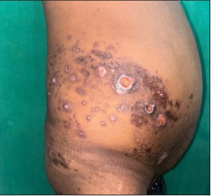
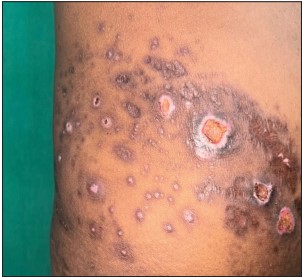
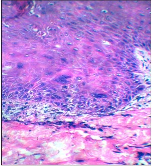
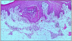
Introduction: Herpes zoster (HZ) is the prototypical cause of zosteriform dermatoses. Additionally, many infectious, inflammatory, and neoplastic conditions may mimic this distribution pattern. Immuno compromised patients, such as those with end-stage renal disease (ESRD), are predisposed to both infectious and reactive skin conditions. In these cases, HZ may present with atypical morphologies, leading to diagnostic challenges. This report presents a case of atypical herpes zoster in a patient with chronic kidney disease (CKD) showing dimorphic ulcerative ecthymatous lesions and crusted umbilicated papulonodules mimicking reactive perforating collagenosis (RPC). This report highlights the importance of histopathology in distinguishing these conditions. Case Report: A 48-year-old male with stage - VCKD on haemodialysis, presented with acute onset of unilateral, multiple painful papulonodules culminating into ulcers over the right lower back and abdomen. The lesions were diverse ranging from punched out ulcersto umbilicated papules with central keratotic crusting. This evoked the differentials of ecthyma or segmental RPC. None of the past or present lesions were vesicular. Investigations revealed anaemia, deranged sugar levels and renal function parameters. Bacterial cultures showed no growth, while Tzanck smear demonstrated multinucleated giant cells. Skin biopsy revealed viral cytopathic changes and follicular involvement typifying herpes viral infection. The patient responded well to renal-adjusted acyclovir therapy, with significant improvement in symptoms and healing of ulcers. Conclusion: Atypical morphologies of herpes zoster, such as ecthymatous and that mirroring RPC, should be considered in immunosuppressed patients. Misdiagnosis as ecthyma or perforating dermatosis can delay appropriate antiviral treatment. Histopathology plays a crucial role in distinguishing HZ from its mimickers. This case underscores the need for heightened clinical suspicion while diagnosing atypical zoster and early intervention to prevent further complications in CKD patients.
References
- 1. Hayderi L., Libon F., Nikkels-Tassoudji N., Rübben A., Dezfoulian B., Nikkels A.F. Zosteriform dermatoses—A review. Glob Dermatol. 2015; 2(4): 163-173. doi:10.15761/ GOD.1000146.
- 2. Yamauchi Y., Oyama N., Hasegawa M. A case of segmental acquired reactive perforating collagenosis: case report and literature review of the unique presentation. An Bras Dermatol. 2025; 100(1): 169-172. doi:10.1016/j. abd.2024.03.010
- 3. Mueller N.H., Gilden D.H., Cohrs R.J., Mahalingam R., Nagel M.A. Varicella-zoster virus infection: clinical features, molecular pathogenesis of disease, and latency. NeurolClin. 2008; 26(3): 675-97, viii.
- 4. Kroth J., Tischer J., Samtleben W., Weiss C., Ruzicka T., et al. Isotopic response, Köbner phenomenon and Renbök phenomenon following herpes zoster. J Dermatol. 2011; 38(11): 1058-61.
- 5. Omidian M., Mapar M.A. Chronic zosteriform cutaneous leishmaniasis. Indian J Dermatol Venereol Leprol. 2006; 72(1): 41-42. doi:10.4103/0378-6323.19717
- 6. Nakanishi G., Tsunemitsu R., Akagi O. Reactive perforating collagenosis occurring in a zosteriform distribution. Br J Dermatol. 1999; 141(2): 367-9.
- 7. Bang S.W., Kim Y.K., Whang K.U. Acquired reactive perforating collagenosis: unilateral umbilicated papules along the lesions of herpes zoster. J Am Acad Dermatol. 1997;36:778-9.
- 8. Lee H.N., Lee D.W., Lee J.Y., Cho B.K. Two cases of reactive perforating collagenosis arising at the site of healed herpes zoster. Int J Dermatol. 2001; 40(3): 191-2. doi:10.1046/ j.1365-4362.2001.01138.x.
- 9. Böer A., Herder N., Blödorn-Schlicht N., Steinkraus V., Falk T.M. Refining criteria for diagnosis of cutaneous infections caused by herpes viruses through correlation of morphology with molecular pathology. Indian J Dermatol Venereol Leprol. 2006; 72(4): 270-5. doi:10.4103/0378-6323.26717.
- 10. Seront E., Kidd F., Metz T., Van Ruyssevelt P. Atypical case of ecthyma gangrenosum mimicking a breast cancer recurrence. BMJ Case Rep. 2016 Mar 30;2016:bcr2016214349. doi:10.1136/bcr-2016-214349.
- 11. Seshadri D., Kumaran M.S., Kanwar A.J. Acantholysis revisited: Back to basics. Indian J Dermatol Venereol Leprol. 2013;79(1):120-6. doi:10.4103/0378-6323.104688.
- 12. Gilson I.H., Barnett J.H., Conant M.A., Laskin OL, Williams J., Jones P.G. Disseminated ecthymatous herpes varicella-zoster virus infection in patients with acquired immunodeficiency syndrome. J Am Acad Dermatol. 1989; 20(4): 637-42. doi:10.1016/ S0190-9622(89)70076-1.
- 13. Schneiderman H., Robert N.J., Walker S., et al. Herpes without vesicles: limited, recurrent genital lesions in an immuno debilitated host. South Med J. 1986; 79(3): 368-70.
- 14. Williamson B.C. Disseminated herpes zoster in a human immunodeficiency virus-positive homosexual man without complications. Cutis. 1987; 40(6): 485-6.
- 15. Walsh N., Boutilier R., Shaffelburg M. Exclusive involvement of folliculosebaceous units by herpes: A reflection of early herpes zoster. Am J Dermatopathol. 2005; 27(3): 189-94.
- 16. Sadjadi S.A., Regmi S., Chau T. Acyclovir neurotoxicity in a peritoneal dialysis patient: report of a case and review of the pharmacokinetics of acyclovir. Am J Case Rep. 2018; 19: 1459-1462. doi:10.12659/AJCR.911520.
Data Sharing Statement
There are no additional data available. All raw data and code are available upon request.
Funding
This research received no funding.
Author Contributions
All authors contributed significantly to the work and approve its publication.
Ethics Declaration
This article does not involve any human or animal subjects, and therefore does not require ethics approval.
Acknowledgements
We would like to express our gratitude to the patients, their families, and all those who have contributed to this study.
Conflicts of Interest
No conflicts of interest in this work.
About this article

Cite this article
Vijay Joshi, Dipti Thakur, Vidya Kharkar. Tale of an Atypical Herpes Zoster Mimicking Ecthyma and Perforating Dermatosis in Chronic Kidney Disease. RFP Jr of Drea 2025; 10(1): 21-27.
Licence:
Attribution-Non-commercial 4.0 International (CC BY-NC 4.0)This license enables reusers to distribute, remix, adapt, and build upon the material in any medium or format for noncommercial purposes only, and only so long as attribution is given to the creator.
| Received | Accepted | Published |
|---|---|---|
| April 23, 2025 | May 12, 2025 | June 25, 2025 |
DOI: https://doi.org/10.21088/jd.2582.3582 10125.4
Keywords
Herpes zosterZosteriform dermatosesEcthymaReactive perforating collagenosisSearch for Similar Articles
Similar Articles
- Delving into Wolf’s Isotopic Phenomenon: Tale of Two Cases of Psoriasis Localize...
- Pachydermoperiostosis, Complete Form: A Case Report of Rare Occurrence
- Role of Cyclical Negative Pressure Wound Therapy in Thermal Burns
- Behcets Disease in an Immunocompromised Female
- Role of Prolotherapy in Wound Bed Preparation in Grade 4 Ischial Pressure Injuri...
Article Level Metrics
Last UpdatedWednesday 21 October 2026, 03:51:16 (IST)
1724
Accesses
13
194
00
Citations
NA
NA
NA
Download citation
Article Keywords
Keyword Highlighting
Highlight selected keywords in the article text.
Timeline
| Received | April 23, 2025 |
| Accepted | May 12, 2025 |
| Published | June 25, 2025 |
licence
Attribution-Non-commercial 4.0 International (CC BY-NC 4.0)
This license enables reusers to distribute, remix, adapt, and build upon the material in any medium or format for noncommercial purposes only, and only so long as attribution is given to the creator.