Full Text (PDF)
Indian Journal of Emergency Medicine 12(3):p 135-138, July - Sept. 2026. | DOI: 10.21088/ijem.2395.311X.12326.2
Case Report
Accidental Retention and Ultrasound-Guided Retrieval of a Fractured IV Cannula During Diagnostic Pleural Tap: A Case Report
Anjani Priya Vamula, G Prasad, Hari Deep Y, Maruthi Rama Krishna, IS Chaitanya Kumar, Muni Srikanth
Author Information
Licence:
Attribution-Non-commercial 4.0 International (CC BY-NC 4.0)This license enables reusers to distribute, remix, adapt, and build upon the material in any medium or format for noncommercial purposes only, and only so long as attribution is given to the creator.
Indian Journal of Emergency Medicine 12(3):p 135-138, July - Sept. 2026. | DOI: 10.21088/ijem.2395.311X.12326.2
How Cite This Article:
Anjani Priya Vamula, G Prasad, Hari Deep et. al, Accidental Retention and Ultrasound-Guided Retrieval of a Fractured IV Cannula During Diagnostic Pleural Tap: A Case Report. Ind J Emerg Med. 2026; 12(3): 135-138.Timeline
Received : May 06, 2026
Accepted : June 09, 2026
Published : September 30, 2026
Abstract
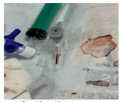
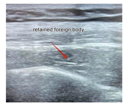
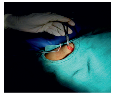
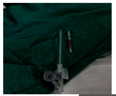
Background: Thoracentesis is a commonly performed diagnostic and therapeutic procedure. Although generally safe, rare complications such as retained foreign bodies may occur. Case Presentation: We describe a case involving a 40-year-old female who presented with fever and progressive dyspnea. Imaging revealed a right-sided pleural effusion. During diagnostic pleural tap using a 16G intravenous (IV) cannula, the catheter fractured, with the proximal two-thirds retained in the chest wall. Immediate point-of-care ultrasound (POCUS) and high-resolution computed tomography (HRCT) localized the fragment. The retained segment was successfully retrieved surgically under ultrasound guidance, and the patient recovered uneventfully. Conclusion: This report highlights the importance of vigilance during pleural procedures and emphasizes the role of multimodal imaging, particularly POCUS and HRCT, in the safe localization and retrieval of retained foreign bodies
References
- 1. Feller-Kopman D. Therapeutic thoracentesis: the role of ultrasound and pleural manometry.
- 2. Al-Qurayshi ZH, Hauch AT, Slakey DP, et al. Retained foreign bodies: risk and outcomes at the national level. J Am Coll Surg. 2015 Apr;220(4):749–759. doi:10.1016/j. jamcollsurg.2014.12.015.
- 3. Husain LF, Hagopian L, Wayman D, et al. Sonographic diagnosis of pneumothorax. Crit
- 4. Scialanga B, Buonsenso D, Scateni S, et al. Lung ultrasound to detect pneumothorax
- 5. Arua OA, Bolaji AA, Ubanatu P, Echieh CP. Retained fractured intravenous catheter:
- 6. Kumar RR, Ranjan P. Case report: Iatrogenic fracture of intravenous cannula during removal with proximal migration. Int J Surg Case Rep. 2020;76:562–565. PMID:33109488.
- 7. Turner DD, Sommers SC. Accidental passage of a polyethylene catheter from cubital vein
Data Sharing Statement
There are no additional data available. All raw data and code are available upon request.
Funding
This research received no funding.
Author Contributions
All authors contributed significantly to the work and approve its publication.
Ethics Declaration
This article does not involve any human or animal subjects, and therefore does not require ethics approval.
Acknowledgements
We would like to express our gratitude to the patients, their families, and all those who have contributed to this study.
Conflicts of Interest
The authors report no conflicts of interest in this work.
About this article

Cite this article
Anjani Priya Vamula, G Prasad, Hari Deep et. al, Accidental Retention and Ultrasound-Guided Retrieval of a Fractured IV Cannula During Diagnostic Pleural Tap: A Case Report. Ind J Emerg Med. 2026; 12(3): 135-138.
Licence:
Attribution-Non-commercial 4.0 International (CC BY-NC 4.0)This license enables reusers to distribute, remix, adapt, and build upon the material in any medium or format for noncommercial purposes only, and only so long as attribution is given to the creator.
| Received | Accepted | Published |
|---|---|---|
| May 06, 2026 | June 09, 2026 | September 30, 2026 |
DOI: 10.21088/ijem.2395.311X.12326.2
Keywords
ThoracentesisFractured IV CannulaPleural EffusionUltrasound-Guided RetrievalSearch for Similar Articles
Similar Articles
- Inherited Thrombophilia as a Cause of Post-Partum Massive Pulmonary Embolism: A...
- ATAK Complex Presenting as Hypertensive Pulmonary Edema Following Meropenem-Indu...
- When Infection Triggers Thrombosis: A Case of Extensive DVT in a Young Male
- Case Report: Diagnostic Dilemma in 69-Year-Old Male with Radiating Right Shoulde...
- The Silent Gallbladder, The Slowing Heart: Cope’s Sign in Acute Calculous Cholec...
Article Level Metrics
Last UpdatedWednesday 21 October 2026, 07:47:59 (IST)
6092
Accesses
19
1017
00
Citations
NA
NA
NA
Download citation
Article Keywords
Keyword Highlighting
Highlight selected keywords in the article text.
Timeline
| Received | May 06, 2026 |
| Accepted | June 09, 2026 |
| Published | September 30, 2026 |
licence
Attribution-Non-commercial 4.0 International (CC BY-NC 4.0)
This license enables reusers to distribute, remix, adapt, and build upon the material in any medium or format for noncommercial purposes only, and only so long as attribution is given to the creator.