Full Text (PDF)
Indian Journal of Genetics and Molecular Research 15(1):p 23-34, Jan-June 2026. | DOI: https://doi.org/10.21088/ijgmr.2319.4782.15126.3
Original Article
A Translational Strategy to Overcome Platinum Resistance in Ovarian Cancer via PARP-1 and GCS Inhibition
Luxmi Devi, Ashok Sharma, Sandeep Mathur, Lalit Kumar
Author Information
Licence:
Attribution-Non-commercial 4.0 International (CC BY-NC 4.0)This license enables reusers to distribute, remix, adapt, and build upon the material in any medium or format for noncommercial purposes only, and only so long as attribution is given to the creator.
Indian Journal of Genetics and Molecular Research 15(1):p 23-34, Jan-June 2026. | DOI: https://doi.org/10.21088/ijgmr.2319.4782.15126.3
How Cite This Article:
Timeline
Received : January 21, 2026
Accepted : February 25, 2026
Published : June 30, 2026
Abstract
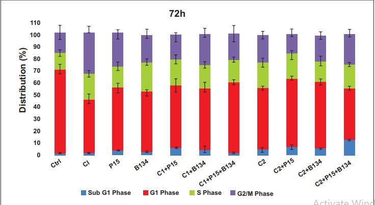
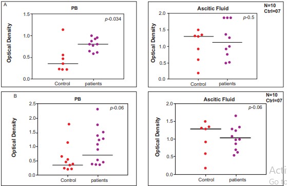
Background: Ovarian cancer (OC) is the most common gynaecological malignancy among women worldwide, with a significant disease burden in India (incidence: 6.6%; mortality: 7.4% per 100,000). Epithelial ovarian cancer (EOC) is the predominant subtype, typically diagnosed at an advanced stage. Standard treatment involves cytoreductive surgery followed by adjuvant chemotherapy with a taxane platinum combination, which initially shows favourable clinical outcomes. However, the majority of patients relapse within 12–18 months due to the development of chemoresistance. Methods: The cytotoxic effects of CDDP, PJ34, and BSO were evaluated individually and in combination using MTT assays in ovarian cancer cell lines (KURAMOCHI, OVSAHO, and IGROV1). Cell cycle and apoptosis were analysed by flow cytometry. Gene expression of PARP-1 and GCS in peripheral blood (PB) and ascitic fluid of newly diagnosed and relapsed ovarian cancer patients was assessed using QRT-PCR, and GSH levels were quantified using ELISA. Results: CDDP showed greater cytotoxicity than PJ34 and BSO, with the IGROV1 cell line exhibiting the highest sensitivity. Combination treatment with CDDP + PJ34 + BSO demonstrated a synergistic effect, significantly reducing cell viability and enhancing cell death compared to monotherapies. Co-treatment induced S-phase arrest and promoted apoptosis. GCS MRNA was significantly upregulated in PB and ascitic fluid of newly diagnosed patients, whereas PARP-1 expression remained unchanged. Elevated GSH levels in PB and reduced levels in ascitic fluid correlated with GCS activity, suggesting a redox imbalance contributing to chemoresistance.
References
- 1. H. Sung et al., “Global cancer statistics 2020: GLOBOCAN estimates of incidence and mortality worldwide for 36 cancers in 185 countries,” CA. Cancer J. Clin., Feb. 2021, doi: 10.3322/caac.21660.
- 2. “Cancer Today.” https://gco.iarc.fr/today/ en/dataviz/pie?mode=cancer&group_populations=1&sexes=2&populations=356&types=0(accessed Feb. 25, 2025).
- 3. I. Ray-Coquard et al., “Non-epithelial ovariancancer: ESMO Clinical Practice Guidelines for diagnosis, treatment and follow-up †,” ESMO Guidel. Comm., vol. 29, pp. iv1–iv18, 2018, doi:10.1093/annonc/mdy001.
- 4. M. McMullen, K. Karakasis, A. Madariaga,and A. M. Oza, “Overcoming platinum and parp-inhibitor resistance in ovarian cancer,” Cancers, vol. 12, no. 6. MDPI AG, pp. 1–18, Jun.01, 2020, doi: 10.3390/cancers12061607.
- 5. C. H. Holschneider and J. S. Berek, “Ovariancancer: Epidemiology, biology, and prognostic factors,” Seminars in Surgical Oncology, vol. 19, no. 1. Semin Surg Oncol, pp. 3–10, Jul. 2000, doi: 10.1002/1098-2388(200007/08)19:1<3::AIDSSU2>3.0.CO;2-S.
- 6. S. Lheureux, C. Gourley, I. Vergote, and A. M.Oza, “Epithelial ovarian cancer,” The Lancet, vol. 393, no. 10177. Lancet Publishing Group, pp. 1240–1253, Mar. 23, 2019, doi: 10.1016/ S0140-6736(18)32552-2.
- 7. S. Shruthi and K. Bhasker Shenoy, “Cisplatin Resistance in Cancer Therapy: Causes and Overcoming Strategies,” ChemistrySelect, vol. 9. no. 25, p. e202401449, Jul. 2024, doi: 10.1002/SLCT.202401449.
- 8. S. W. Johnson, R. F. Ozols, and T. C. Hamilton,“Mechanisms of drug resistance in ovarian cancer,” Cancer, vol. 71, no. 2 S, pp. 644–649, 1993, doi: 10.1002/cncr.2820710224.
- 9. B. Stordal and M. Davey, “Understanding cisplatin resistance using cellular models,” IUBMB Life, vol. 59, no. 11. pp. 696–699, 2007, doi: 10.1080/15216540701636287.
- 10. A. Sharma et al., “Epigenetic activation of POTE genes in ovarian cancer,” Epigenetics, vol. 14, no. 2, p. 185, Feb. 2019, doi:10.1080/15592294.2019.1581590.
- 11. M. Mann et al., “PARP-1 inhibitor modulate β-catenin signaling to enhance cisplatin sensitivity in cancer cervix,” Oncotarget, vol. 10, no. 42, p. 4262, 2019, doi: 10.18632/Oncotarget.27008.
- 12. “Protocol for Phenol/Chloroform RNA Extraction | EpiGentek.” https://www.epigentek.com/catalog/protocol-forphenolchloroform-rna-extraction-n-43.html? news Path=1 (accessed Jul. 22, 2021).
- 13. A. Tyagi, R. Pramanik, R. Bakhshi, A. Singh,S. Vishnubhatla, and S. Bakhshi, “Expression of mitochondrial genes predicts survival in pediatric acute myeloid leukemia,” Int. J. Hematol., vol. 110, no. 2, pp. 205–212, Aug. 2019, doi: 10.1007/S12185-019-02666-2.
- 14. H. Alizadeh, P. Akbarabadi, A. Dadfar, M. R. Tareh, and B. Soltani, “A comprehensive overview of ovarian cancer stem cells:correlation with high recurrence rate, underlying mechanisms, and therapeutic opportunities,” Mol. Cancer 2025 241, vol. 24,P no. 1, pp. 135-, May 2025, doi: 10.1186/S12943-025-02345-3.
- 15. L. Goenka et al., “Targeting autophagy in platinum-sensitive relapsed ovarian cancer: randomized phase II trial of hydroxychloroquine with chemotherapy with biomarker correlation,” Discov. Oncol. 2025 161, vol. 16, no. 1, pp. 203-, Feb. 2025, doi: 10.1007/S12672-025-01904-W.
- 16. T. Zaremba and N. J. Curtin, “PARP Inhibitor Development for Systemic Cancer Targeting,” Anticancer. Agents Med. Chem., vol. 7, no. 5, pp. 515–523, Sep. 2007, doi: 10.2174/187152007781668715.
- 17. “PARP inhibitors in breast cancer - PubMed.” https://pubmed.ncbi.nlm.nih.gov/21157412/ (accessed May 02, 2021).
- 18. D. Davar, J. H. Beumer, L. Hamieh, and H. Tawbi, “Role of PARP Inhibitors in Cancer Biology and Therapy,” Curr. Med. Chem., vol. 19, no. 23, pp. 3907–3921, Jul. 2012, doi: 10.2174/092986712802002464.
- 19. “PJ34, an inhibitor of PARP-1, suppresses cell growth and enhances the suppressive effects of cisplatin in liver cancer cells - PubMed.” https://pubmed.ncbi.nlm.nih.gov/18695907/ (accessed Jan. 13, 2022).
- 20. G. Noël, C. Godon, M. Fernet, N. Giocanti, F. Mégnin-Chanet, and V. Favaudon, “Radiosensitization by the poly(ADPribose) polymerase inhibitor 4-amino-1,8- naphthalimide is specific of the S phase of the cell cycle and involves arrest of DNA synthesis,” Mol. Cancer Ther., vol. 5, no. 3, pp. 564–574, Mar. 2006, doi: 10.1158/1535-7163. MCT-05-0418.
- 21. M. Cohen-Armon, “The Modified Phenanthridine PJ34 Unveils an Exclusive Cell-Death Mechanism in Human Cancer Cells,” Cancers (Basel)., vol. 12, no. 6, pp. 1–15, Jun. 2020, doi: 10.3390/ Cancers12061628.
- 22. K. S. Lee et al., “Effects of buthionine sulfoximine treatment on cellular glutathione levels and cytotoxicities of cisplatin, carboplatin and radiation in human stomach and ovarian cancer cell lines.,” Korean J. Intern. Med., vol. 7, no. 2, pp. 111–117, 1992, doi: 10.3904/kjim.1992.7.2.111.
- 23. Y. Huang et al., “A GSH-responsive oxidative stress nanoamplifier for self-augmented chemo/chemodynamic therapy to reverse cisplatin resistance,” Acta Biomater., vol. 193, pp. 440–454, Feb. 2025, doi: 10.1016/J. ACTBIO.2024.12.041.
- 24. Q. Li et al., “The effects of buthionine sulfoximine on the proliferation and apoptosis of biliary tract cancer cells induced by cisplatin and gemcitabine,” Oncol. Lett., vol. 11, no. 1, pp. 474–480, Jan. 2016, doi: 10.3892/ol.2015.3879.
- 25. W. Liang, Y. Huang, Y. Wang, D. Lu, and Q. Sun, “Research Progress of PlatinumBased Complexes in Lung Cancer Treatment: Mechanisms, Applications, and Challenges,” Int. J. Mol. Sci. 2025, Vol. 26, Page 7958, vol. 26, no. 16, p. 7958, Aug. 2025, doi: 10.3390/IJMS26167958.
- 26. S.M et al., “Enhanced polyadenosine diphosphate-ribosylation in cirrhotic liver and carcinoma tissues in patients with hepatocellular carcinoma,” J. Gastroenterol. Hepatol., vol. 16, no. 3, pp. 338–344, 2001, doi:10.1046/J.1440-1746.2001.02378.X.
- 27. Y.L, T.S.L, S.A, Y.G, T.D, and B.E, “Changes in NAD/ADP-ribose metabolism in rectal cancer,” Brazilian J. Med. Biol. Res. = Rev. Bras. Pesqui. medicas e Biol., vol. 38, no.3, pp. 361–365, 2005, doi: 10.1590/S0100-879X2005000300006.
- 28. T. Borggrefe, M. Wabl, A.T. Akhmedov, and R. Jessberger, “A B-cell-specific DNA recombination complex,” J. Biol. Chem., vol.273, no. 27, pp. 17025–17035, Jul. 1998, doi:10.1074/jbc.273.27.17025.
- 29. “Poly(ADP-ribose) polymerase gene expression status and genomic instability in human breast cancer - PubMed.” https://pubmed.ncbi.nlm.nih.gov/9816283/ (accessed Jun. 01, 2021).
- 30. J. Kigawa, Y. Minagawa, X. Cheng, and N. Terakawa, “yy Glutamyl Cysteine Synthetase Up-Regulates Glutathione and Multidrug Resistance-associated Protein in Patients with Chemoresistant Epithelial Ovarian Cancer,” 1998.
- 31. K.S. Yao, A.K. Godwin, S.W. Johnson, R.F. Ozols, P.J. O’dwyer, and T.C. Hamilton, “Evidence for Altered Regulation of y-Glutamylcysteine Synthetase Gene Expression among Cisplatinsensitive and Cisplatin-resistant Human Ovarian Cancer Cell Lines1,” 1995.
- 32. S. Ullah Khan, “Connotation of GlutathioneS-Transferase Enzyme Expression (GST) and Glutathione (GSH) in Human Head and Neck Squamous Cell Carcinoma Cancer,” J. Cancer Prev. Curr. Res., vol. 2, no. 5, May 2015, doi: 10.15406/jcpcr.2015.02.00051.
Data Sharing Statement
There are no additional data available. All raw data and code are available upon request.
Funding
This research received no funding.
Author Contributions
All authors contributed significantly to the work and approve its publication
Ethics Declaration
This article does not involve any human or animal subjects, and therefore does not require ethics approval.
Acknowledgements
We would like to express our gratitude to the patients, their families, and all those who have contributed to this study
Conflicts of Interest
No conflicts of interest in this work
About this article
Cite this article
Licence:
Attribution-Non-commercial 4.0 International (CC BY-NC 4.0)This license enables reusers to distribute, remix, adapt, and build upon the material in any medium or format for noncommercial purposes only, and only so long as attribution is given to the creator.
| Received | Accepted | Published |
|---|---|---|
| January 21, 2026 | February 25, 2026 | June 30, 2026 |
DOI: https://doi.org/10.21088/ijgmr.2319.4782.15126.3
Keywords
Ovarian CancerCisplatinPARP-1PJ34Y-GCSB-actinSearch for Similar Articles
Similar Articles
- Success After Unexplained Multiple Miscarriages using PGT-A and PGT-M for RELN G...
- Bridging Classic Cytogenetics with Modern Chromosomics: A Historical and Scienti...
- Mitochondrial D-Loop and HBB Gene Profiling in Antenatal Hemoglobinopathy Carrie...
- Histopathologic Features of High Grade Serous Ovarian Carcinomas Associated with...
- Genetic Insight into Familial and Sporadic Alzheimer’s Disease
Article Level Metrics
Last UpdatedThursday 30 July 2026, 07:16:09 (IST)
1248
Accesses
23
313
00
Citations
NA
NA
NA
Download citation
Article Keywords
Keyword Highlighting
Highlight selected keywords in the article text.
Timeline
| Received | January 21, 2026 |
| Accepted | February 25, 2026 |
| Published | June 30, 2026 |
licence
Attribution-Non-commercial 4.0 International (CC BY-NC 4.0)
This license enables reusers to distribute, remix, adapt, and build upon the material in any medium or format for noncommercial purposes only, and only so long as attribution is given to the creator.