Full Text (PDF)
Indian Journal of Emergency Medicine 10 (4):p 248-251, October - December 2024. | DOI: https://doi.org/10.21088/ijem.2395.311X.10424.10
Case Report
A Case Report of Spinal Vascular Malformation as a Cause of Progressive Myelopathy in a 32-Year-Old Female
Renaldo Pavrey, Nikita Makwana
Author Information
Licence:
Attribution-Non-commercial 4.0 International (CC BY-NC 4.0)This license enables reusers to distribute, remix, adapt, and build upon the material in any medium or format for noncommercial purposes only, and only so long as attribution is given to the creator.
Indian Journal of Emergency Medicine 10 (4):p 248-251, October - December 2024. | DOI: https://doi.org/10.21088/ijem.2395.311X.10424.10
How Cite This Article:
34. Pavrey R, Makwana N. A case report of spinal vascular malformation as a cause of progressive myelopathy in a 32-year-old female. Ind J Emerg Med. 2024;10(4):248-51.Timeline
Received : July 04, 2024
Accepted : August 14, 2024
Published : December 15, 2024
Abstract
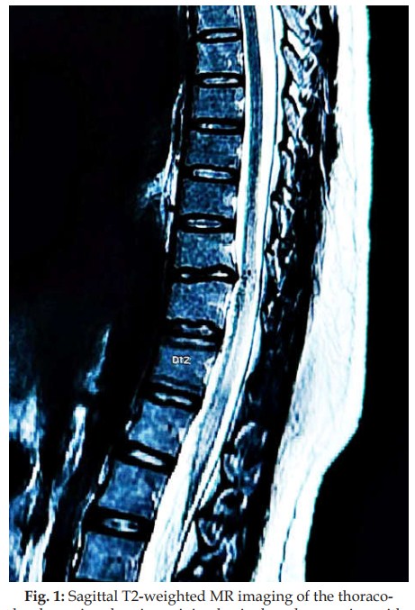
Spinal arteriovenous malformations (AVMs) are a rare form of spinal blood vessel defect that result in engorgement of the vessel leading to clinical manifestations due to mass effect and cord ischemia. They are a heterogeneous group that can cause acute, subacute, or chronic spinal cord dysfunction. Most of these patients present to the Emergency Department (ED) after a prolonged course with severe neurological disability. Spinal vascular lesions comprise approximately 3-4 % of all intradural spinal lesions. When it comes to clinical impact, these lesions have a comparatively worse curve when compared to their intracranial counterparts. We describe the case of a 32-year-old female with no previous medical co-morbid conditions, who presented to us with the symptoms of progressive bilateral lower limb weakness since 3 weeks, with the subsequent inability to stand. There was no urinary bladder or bowel involvement. Her physical examination was positive for lower truncal and lower limb proximal muscle weakness (left > right), with diminished sensations corresponding to the T9-T10 dermatome levels. Magnetic resonance imaging (MRI) of the thoraco-lumbar spine showed a type IV vascular malformation in the left anterior perimedullary region of T10 vertebra spanning to L1. Unfortunately, the patient left the Emergency Department against medical advice, due to severe financial constraints, and was lost to follow-up. We believe this case to be of clinical value because intradural perimedullary arteriovenous fistula are rare, with true incidence rates not yet defined. Our aim is to highlight this extremely uncommon cause of myelopathy presenting as progressive paraparesis in the Emergency Department.
References
- 1. Lad SP, Santarelli JG, Patil CG, Steinberg GK, Boakye M. National trends in spinal arteriovenous malformations. Neurosurg Focus. 2009; 26:1–5.
- 2. Patchana T, Savla P, Taka TM, Ghanchi H, Wiginton J 4th, Schiraldi M, Cortez V. Spinal Arteriovenous Malformation: Case Report and Review of the Literature. Cureus. 2020 Nov 21; 12(11):e11614. doi: 10.7759/cureus.11614. PMID: 33364131; PMCID: PMC7752798.
- 3. Di Chiro G, Doppman J, Ommaya A. Selective Arteriography of Arteriovenous Aneurysms of Spinal Cord. Radiology. 1967; 88(6):1065-77. doi:10.1148/88.6.1065.
- 4. Takai K. Spinal Arteriovenous Shunts: Angioarchitecture and Historical Changes in Classification. Neurol Med Chir (Tokyo). 2017; 57(7):356-65. doi:10.2176/nmc.ra.2016-0316.
- 5. da Costa L, Dehdashti AR, terBrugge KG. Spinal cord vascular shunts: spinal cord vascular malformations and dural arteriovenous fistulas. Neurosurg Focus. 2009; 26:E6.
- 6. Krings T, Mull M, Gilsbach JM, Thron A. Spinal vascular malformations. EurRadiol. 2005; 15:267–278.
- 7. Rosenblum B, Oldfield EH, Doppman JL, Di Chiro G. Spinal arteriovenous malformations: a comparison of dural arteriovenous fistulas and intradural AVM’s in 81 patients. J Neurosurg. 1987; 67:795–802.
- 8. Gueguen B, Merland JJ, Riche MC, Rey A. Vascular malformations of the spinal cord: intrathecal perimedullary arteriovenous fistulas fed by medullary arteries. Neurology. 1987;37:969–979.
- 9. Grote EH, Voigt K. Clinical syndromes, natural history, and pathophysiology of vascular lesions of the spinal cord. NeurosurgClin N Am. 1999; 10:17–45.
- 10. Gross BA, Du R. Spinal glomus (type II) arteriovenous malformations: a pooled analysis of hemorrhage risk and results of intervention. Neurosurgery. 2013; 72:25–32.
- 11. Jeng Y, Chen DY-T, Hsu H-L, Huang Y-L, Chen C-J, Tseng Y-C. Spinal dural arteriovenous fistula: imaging features and its mimics. Korean J Radiol. 2015; 16:1119–1131.
- 12. Murai S, Hiramatsu M, Suzuki E et al. Trends in Incidence of Intracranial and Spinal Arteriovenous Shunts: HospitalBased Surveillance in Okayama, Japan. Stroke. 2021; 52(4):1455 – 1459.doi: 10.1161/ STROKEAHA.120.032052.
- 13. Ozpinar A, Weiner GM, Ducruet AF. Epidemiology, clinical presentation, diagnostic evaluation, and prognosis of spinal arteriovenous malformations. In:Spetzler RF, Moon K, Almefty RO, editors. Handbook of Clinical Neurology. Vol. 143. Netherlands: Elsevier; 2017. 145-52.
- 14. Jellema K, Canta LR, Tijssen CC, van Rooij WJ, Koudstaal PJ, van Gijn J. Spinal dural arteriovenous fistulas:clinical features in 80 patients. J NeurolNeurosurg Psychiatry. 2003; 74:1438-40.
- 15. Nagashima C, Miyoshi A, Nagashima R, Ogawa M, Enomoto K, Watabe T. Spinal giant intradural perimedullary arteriovenous fistula: clinical and neuroradiological study in one case with review of literature.Surg Neurol. 1996; 45:524–531.
Data Sharing Statement
There are no additional data available.
Funding
This research received no funding.
Author Contributions
All authors contributed significantly to the work and approve its publication.
Acknowledgements
Information not provided.
Conflicts of Interest
The authors declare no conflicts of interest regarding the publication of this paper.
About this article
Cite this article
34. Pavrey R, Makwana N. A case report of spinal vascular malformation as a cause of progressive myelopathy in a 32-year-old female. Ind J Emerg Med. 2024;10(4):248-51.
Licence:
Attribution-Non-commercial 4.0 International (CC BY-NC 4.0)This license enables reusers to distribute, remix, adapt, and build upon the material in any medium or format for noncommercial purposes only, and only so long as attribution is given to the creator.
| Received | Accepted | Published |
|---|---|---|
| July 04, 2024 | August 14, 2024 | December 15, 2024 |
DOI: https://doi.org/10.21088/ijem.2395.311X.10424.10
Keywords
spinal vascular malformationArteriovenous fistulaMyelopathySearch for Similar Articles
Similar Articles
- Crossroads of Industrial Chemistry and Clinical Toxicology: A Rare Case of Orga...
- A Tale of Double Vision
- Incidental Diagnosis of Graves’ Disease in a Patient Presenting with Influenza:...
- Avulsion Fracture Hidden in X-Ray Detected by Point-of-Care Ultrasound in a Youn...
- Disseminated Deep Neck Space Infection in an Immunocompetent Adult: A Case Repor...
Article Level Metrics
Last UpdatedMonday 22 June 2026, 03:49:50 (IST)
2182
Accesses
8
767
00
Citations
NA
NA
NA
Download citation
Article Keywords
Keyword Highlighting
Highlight selected keywords in the article text.
Timeline
| Received | July 04, 2024 |
| Accepted | August 14, 2024 |
| Published | December 15, 2024 |
licence
Attribution-Non-commercial 4.0 International (CC BY-NC 4.0)
This license enables reusers to distribute, remix, adapt, and build upon the material in any medium or format for noncommercial purposes only, and only so long as attribution is given to the creator.