Full Text (PDF)
Indian Journal of Emergency Medicine 12(1):p 39-43, Jan. March 2026. | DOI: https://doi.org/10.21088/ijem.2395.311X.12126.7
Case Report
Avulsion Fracture Hidden in X-Ray Detected by Point-of-Care Ultrasound in a Young Athlete
Sreelakshmi K.T, Sangeeta Sao, Priya Govil, Kishalay Datta
Author Information
Licence:
Attribution-Non-commercial 4.0 International (CC BY-NC 4.0)This license enables reusers to distribute, remix, adapt, and build upon the material in any medium or format for noncommercial purposes only, and only so long as attribution is given to the creator.
Indian Journal of Emergency Medicine 12(1):p 39-43, Jan. March 2026. | DOI: https://doi.org/10.21088/ijem.2395.311X.12126.7
How Cite This Article:
Sreelakshmi K.T., Sangeeta Sao, Priya Govil, et al. Avulsion Fracture Hidden in X-Ray Detected by Point-of Care Ultrasound in a Young Athlete. Ind J Emerg Med. 2026; 12(1): 39-43.Timeline
Received : July 18, 2025
Accepted : September 05, 2025
Published : March 30, 2026
Abstract
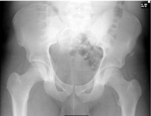
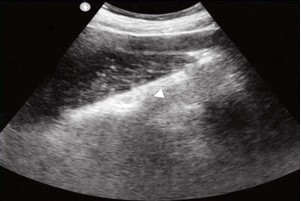
Anterior superior iliac spine (ASIS) avulsion fractures are uncommon injuries, primarily affecting adolescent athletes during sudden, forceful physical activity. Traditional diagnosis relies on radiography, though small bony fragments can be missed. We report a case of a 15-year-old female presenting with acute left groin pain following a rapid directional change while playing “kho-kho”. Physical examination showed localized tenderness, and estricted hip movement. Pelvic X-ray unfortunately skipped the diagnosis when the Point-of-care ultrasound (POCUS) performed in the emergency department revealed a hyperechoic apophyseal fragment displaced from the ASIS, suggesting an avulsion injury. Given minimal displacement, the patient was managed conservatively. This case highlights the value of POCUS in Indian emergency settings where quick decision making and limited imaging facilities make bedside diagnosis essential
References
- 1. Schiller J., DeFroda S., Blood T. Lower Extremity Avulsion Fractures in the Pediatric and Adolescent Athlete. J Am Acad Orthop Surg. 2017; 25(4): 251-9. https://doi.org/10.5435/ JAAOS-D-15-00328.
- 3. Serbest S., Tosun H.B., Tiftikci U., Oktas B., Kesgin E. Anterior inferior iliac spine avulsion fracture: a series of 5 cases. Medicine (Baltimore). 2015; 94(7): e562. https://doi. org/10.1097/MD.0000000000000562.
- 4. Rossi F., Dragoni S. Acute avulsion fractures of the pelvis in adolescent competitive athletes: prevalence, location and sports distribution of 203 cases collected. Skeletal Radiol. 2001; 30(3): 127-31. https://doi.org/10.1007/s002560000319. Figure 4. X-ray of the pelvis. A slightly displaced radiopaque structure was detected on the lateral side of the ASIS, suggesting an ASIS avulsion fracture (arrow). 143 | POCUS J | APR 2022 vol. 07 iss. 01
- 7. Jones S., Colaco K., Fischer J., Stimec J., Kwan C., Boutis K. Accuracy of Point-of Care Ultrasonography for Pediatric Ankle Sprain Injuries. Pediatr Emerg Care. 2018; 34(12): 842-7. https://doi.org/10.1097/ PEC.0000000000001130.
- 8. Chen L.W., Yeh W.C. Musculoskeletal Sonography Facilitates the Diagnosis of Adolescent Anterior Superior Iliac Spine Avulsion Fracture. Journal of Medical Ultrasound. 2010;18(4):158-60. https://doi. org/10.1016/j.jmu.2010.11.003.
- 10. Martinoli C, Valle M, Malattia C, Beatrice Damasio M, Tagliafico A. Paediatric musculoskeletal US beyond the hip joint. Pediatr Radiol. 2011; 41 Suppl 1: S113-24. https://doi.org/10.1007/s00247-011-2037-z.
- 11. Koulouris G., Connell D. Evaluation of the hamstring muscle complex following acute injury. Skeletal Radiol. 2003; 32(10): 582-9. https://doi.org/10.1007/s00256-003-0674-5.
- 12. Marin J.R., Abo A.M., Arroyo A.C., Doniger S.J., Fischer J.W., Rempell R., et al. Pediatric emergency medicine point-of-care ultrasound: summary of the evidence. Crit Ultrasound J. 2016; 8(1): 16. https://doi.org/10.1186/ s13089-016-0049-5.
- 13. Zhang B.F., Lei J.L., Zhang H., Wang P.F., Wang H., Cong Y.X., et al. Use of ultrasonography for evaluation of stability of lateral compression type 1 (LC-1) pelvic fractures to assist determination of treatment strategy. J Orthop Surg Res. 2019; 14(1): 7. https://doi. org/10.1186/s13018-018-1047-z.
- 14. Chartier L.B., Bosco L., Lapointe-Shaw L., Chenkin J. Use of point-ofcare ultrasound IJEM/Volume 12 Number 1/January–March 2026 Sreelakshmi K.T., Sangeeta Sao, Priya Govil, et al. Avulsion Fracture Hidden in X-Ray Detected by Point-of-Care Ultrasound in a Young Athlete. in long bone fractures: A systematic review and metaanalysis. CJEM. 2017; 19(2): 131-42. https://doi.org/10.1017/cem.2016.397.View
- 15. Pisacano R.M., Miller T.T. Comparing sonography with MR imaging of apophyseal 43 injuries of the pelvis in four boys. AJR Am J Roentgenol. 2003; 181(1): 223-30. https://doi. org/10.2214/ajr.181.1.1810223.
Funding
This research received no funding.
Author Contributions
All authors contributed significantly to the work and approve its publication.
Ethics Declaration
This article does not involve any human or animal subjects, and therefore does not require ethics approval.
Acknowledgements
We would like to express our gratitude to the patients, their families, and all those who have contributed to this study.
Conflicts of Interest
No conflicts of interest in this work.
About this article
Cite this article
Sreelakshmi K.T., Sangeeta Sao, Priya Govil, et al. Avulsion Fracture Hidden in X-Ray Detected by Point-of Care Ultrasound in a Young Athlete. Ind J Emerg Med. 2026; 12(1): 39-43.
Licence:
Attribution-Non-commercial 4.0 International (CC BY-NC 4.0)This license enables reusers to distribute, remix, adapt, and build upon the material in any medium or format for noncommercial purposes only, and only so long as attribution is given to the creator.
| Received | Accepted | Published |
|---|---|---|
| July 18, 2025 | September 05, 2025 | March 30, 2026 |
DOI: https://doi.org/10.21088/ijem.2395.311X.12126.7
Keywords
POCUSAvulsion fractureASISkho-khoSearch for Similar Articles
Similar Articles
- Covid 19 Vaccine: Associated Henoch Schonlein Purpura in Young Adult Female
- A Rare Case of Intolerance to Medically Approved Dose of Sildenafil Citrate in a...
- Acute Mesenteric Ischemia: An Unusual Cause of Hematemesis in a 48 Year Old Man
- Brachial Artery Pseudoaneurysm Related Causally to Iatrogenic Coagulopathy in a...
- Combination Therapy for Acute Management of Yellow Oleander Poisoning: A Case Re...
Article Level Metrics
Last UpdatedMonday 27 July 2026, 12:28:28 (IST)
3262
Accesses
24
910
00
Citations
NA
NA
NA
Download citation
Article Keywords
Keyword Highlighting
Highlight selected keywords in the article text.
Timeline
| Received | July 18, 2025 |
| Accepted | September 05, 2025 |
| Published | March 30, 2026 |
licence
Attribution-Non-commercial 4.0 International (CC BY-NC 4.0)
This license enables reusers to distribute, remix, adapt, and build upon the material in any medium or format for noncommercial purposes only, and only so long as attribution is given to the creator.