Full Text (PDF)
Indian Journal of Dental Education 18(3):p 157-160, Sept - Dec. 2025. | DOI: https://doi.org/10.21088/ijde.0974.6099.18325.7
Case Report
Unusual Root Canal Morphology in a Mandibular Second Premolar: A Case Report of a Three-Canal Variation
RVSSK Kinneresh,, D. Karishma, Vajrala Deepika, Sasi Priyanka
Author Information
Licence:
Attribution-Non-commercial 4.0 International (CC BY-NC 4.0)This license enables reusers to distribute, remix, adapt, and build upon the material in any medium or format for noncommercial purposes only, and only so long as attribution is given to the creator.
Indian Journal of Dental Education 18(3):p 157-160, Sept - Dec. 2025. | DOI: https://doi.org/10.21088/ijde.0974.6099.18325.7
How Cite This Article:
D. Karishma, Vajrala Deepika, D. Sreenija, et al. Unusual Root Canal Morphology in a Mandibular Second Premolar: A Case Report of a Three-Canal Variation. Ind J Dent Educ. 2025; 18(3): 157-160.Timeline
Received : June 07, 2025
Accepted : July 10, 2025
Published : December 30, 2025
Abstract
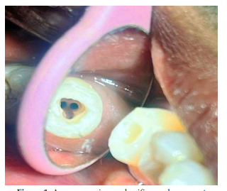
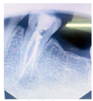
The mandibular second premolar is typically described as having a single root and canal, with anatomical variations being relatively rare. The presence of three distinct canals in this tooth is an uncommon clinical finding that can pose diagnostic and therapeutic challenges. This report presents the successful endodontic management of a mandibular second premolar with three separate root canals. Emphasis is placed on the role of advanced diagnostic techniques, such as angled radiography and careful exploration under magnification, in identifying aberrant canal configurations. A concise review of the prevalence, classification, and clinical implications of such variations is also presented.
References
- 1. Vertucci F.J. Root canal anatomy of the human permanent teeth. Oral Surg Oral Med Oral Pathol. 1984; 58(5): 589–599.
- 2. Cleghorn B.M., Christie W.H., Dong C.C. Root and root canal morphology of the human permanent mandibular second premolar: a literature review. J Endod. 2007; 33(9): 1031–1037.
- 3. Hoen M.M., Pink F.E. Contemporary endodontic retreatments: an analysis based on clinical treatment findings. J Endod. 2002; 28(12): 834–836.
- 4. Slowey R.R. Root canal anatomy: road map to successful endodontics. Dent Clin North Am. 1979; 23(4): 555–573.
- 5. Vertucci F.J. Root canal morphology and its relationship to endodontic procedures. Endod Topics. 2005; 10(1): 3–29.
- 6. Siqueira J.F. Jr. Aetiology of root canal treatment failure: why well-treated teeth can fail. Int Endod J. 2001; 34(1): 1–10.
- 7. Zillich R., Dowson J. Root canal morphology of mandibular first and second premolars. Oral Surg Oral Med Oral Pathol. 1973; 36(5): 738–744.
- 8. Patel S., Durack C., Abella F., Shemesh H., Roig M., Lemberg K. Cone beam computed tomography in Endodontics – A review. Int Endod J. 2015; 48(1): 3–15.
- 9. Trope M., Elfenbein L., Tronstad L. Mandibular premolars with more than one root canal in different race groups. J Endod. 1986; 12(8): 343–345.
- 10. Baldassari-Cruz L.A., Lilly J.P., Rivera E.M. The influence of dental operating microscopes in locating the mesiolingual canal orifice. Oral Surg Oral Med Oral Pathol Oral Radiol Endod. 2002; 93(2): 190–194.
- 11. Buhrley L.J., Barrows M.J., BeGole E.A., Wenckus C.S. Effect of magnification on locating the MB2 canal in maxillary molars. J Endod. 2002; 28(4): 324–327.
- 12. Nair P.N. Pathogenesis of apical periodontitis and the causes of endodontic failures. Crit Rev locating the MB2 canal in maxillary molars. J Endod. 2002; 28(4): 324–327.
Data Sharing Statement
There are no additional data available. All raw data and code are available upon request.
Funding
This research received no funding.
Author Contributions
All authors contributed significantly to the work and approve its publication.
Ethics Declaration
This article does not involve any human or animal subjects, and therefore does not require ethics approval.
Acknowledgements
We would like to express our gratitude to the patients, their families, and all those who have contributed to this study.
Conflicts of Interest
No conflicts of interest in this work.
About this article
Cite this article
D. Karishma, Vajrala Deepika, D. Sreenija, et al. Unusual Root Canal Morphology in a Mandibular Second Premolar: A Case Report of a Three-Canal Variation. Ind J Dent Educ. 2025; 18(3): 157-160.
Licence:
Attribution-Non-commercial 4.0 International (CC BY-NC 4.0)This license enables reusers to distribute, remix, adapt, and build upon the material in any medium or format for noncommercial purposes only, and only so long as attribution is given to the creator.
| Received | Accepted | Published |
|---|---|---|
| June 07, 2025 | July 10, 2025 | December 30, 2025 |
DOI: https://doi.org/10.21088/ijde.0974.6099.18325.7
Keywords
Root Canal MorphologyMandibular Second PremolarAberrant CanalSearch for Similar Articles
Similar Articles
- Post Extraction Surgical Ciliated Cyst of Maxilla: Case Report of Unusual Pathog...
- Natural Tooth Surgical Stent (NTSS) Technique For Prosthodontically Driven Rehab...
- Growth Meets Diagnosis: Revisiting Synchondrosis in the Orthodontic Era
- The Silent Struggle to Breathe: A Review on Obstructive Sleep Apnea
- Evaluating Awareness of Oral-Systemic Health Interactions in Urbanized Indian So...
Article Level Metrics
Last UpdatedTuesday 28 July 2026, 22:29:06 (IST)
1875
Accesses
11
467
00
Citations
NA
NA
NA
Download citation
Article Keywords
Keyword Highlighting
Highlight selected keywords in the article text.
Timeline
| Received | June 07, 2025 |
| Accepted | July 10, 2025 |
| Published | December 30, 2025 |
licence
Attribution-Non-commercial 4.0 International (CC BY-NC 4.0)
This license enables reusers to distribute, remix, adapt, and build upon the material in any medium or format for noncommercial purposes only, and only so long as attribution is given to the creator.