Full Text (PDF)
International Physiology 10(2):p 45-49, May-August 2022. | DOI: https://doi.org/10.21088/ip.2347.1506.10222.4
Original Article
Role of Video Dermatoscopy in Scar Management
Ravi Kumar Chittoria, Gopikrishna Mohanraj, Jacob Antony Chakiath
Author Information
Licence:
Attribution-Non-commercial 4.0 International (CC BY-NC 4.0)This license enables reusers to distribute, remix, adapt, and build upon the material in any medium or format for noncommercial purposes only, and only so long as attribution is given to the creator.
International Physiology 10(2):p 45-49, May-August 2022. | DOI: https://doi.org/10.21088/ip.2347.1506.10222.4
How Cite This Article:
Gopikrishna Mohanraj, Ravi Kumar Chittoria, Jacob Antony Chakiath/Role of Video Dermatoscopy in Scar Management/ International Physiology.2022;10(2):45–49.Timeline
Received : May 30, 2022
Accepted : June 02, 2022
Published : August 25, 2022
Abstract
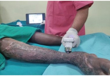
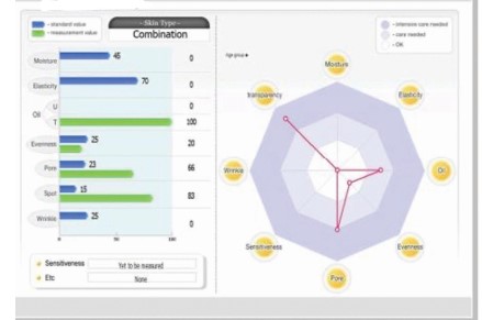
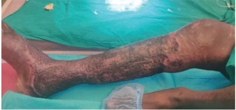
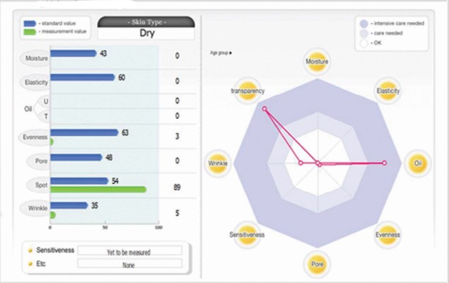
Video Dermatoscopy is a non-invasive method that allows the in-vivo evaluation of colours and microstructures of the epidermis, the dermo-epidermal junction, and the papillary dermis not visible to the naked eye using digital device. This technique is useful for clinician to assess pigmentory/non-pigmentory skin lesion, skin malignancies and skin infections. But usage of Video Dermatoscopy in scar management has scanty data. In this case report we are sharing our experience of Video Dermatoscopy role in scar management.
References
- 1. English RS, Shenefelt PD. Keloids and hypertrophic scars. Dermatol Surg. 1999;25:631–638.
- 2. Campos-do-Carmo G, Ramos-e-Silva M. Dermoscopy: basic concepts. Int J Dermatol. 2008;47:712–719.
- 3. Stolz, Wilhelm (1989).
- 4.
- 5. Hypertrophic scars and keloids: overview of the evidence and practical guide for differentiating between these abnormal scarsG.C. Limandjaja1, F.B. Niessen2, R. Scheper3, S. Gibbs.
- 6. Intraobserver agreement in interpretation of digital epiluminescence microscopy IgnazioStanganelli, MD,a Marco Burroni, PhD,b Silvia Rafanelli, MD,a and Laura Bucchi, MD’ Ravenna, Siena, and Forli, Italy.
- 7. Computerized digital dermoscopy A J Gewirtzman& R P Braun Pigmented Skin Lesion Clinic, Department of Dermatology, University Hospital Geneva, Switzerland.
- 8. Digital dermoscopy in clinical practise: a threecentre analysis Uwe Wollina1, Marco Burroni2, Rocco Torricelli3, Stefano Gilardi4, Giordana Dell’Eva5, Cathrine Helm1 and Wolfgang Bardey.
- 9. Baryza MJ, Baryza GA. The Vancouver Scar Scale: an administration tool and its interrater reliability. J Burn Care Rehabil. 1995;16:535–538.
Data Sharing Statement
There are no additional data available. All raw data and code are available upon request.
Funding
This research received no funding.
Author Contributions
All authors contributed significantly to the work and approve its publication.
Ethics Declaration
This article does not involve any human or animal subjects, and therefore does not require ethics approval.
Acknowledgements
We would like to express our gratitude to the patients, their families, and all those who have contributed to this study.
Conflicts of Interest
No conflicts of interest in this work.
About this article

Cite this article
Gopikrishna Mohanraj, Ravi Kumar Chittoria, Jacob Antony Chakiath/Role of Video Dermatoscopy in Scar Management/ International Physiology.2022;10(2):45–49.
Licence:
Attribution-Non-commercial 4.0 International (CC BY-NC 4.0)This license enables reusers to distribute, remix, adapt, and build upon the material in any medium or format for noncommercial purposes only, and only so long as attribution is given to the creator.
| Received | Accepted | Published |
|---|---|---|
| May 30, 2022 | June 02, 2022 | August 25, 2022 |
DOI: https://doi.org/10.21088/ip.2347.1506.10222.4
Keywords
ScarVideo Dermatoscopy and Assessment toolSearch for Similar Articles
Similar Articles
- Reconstruction of Amputated Stump with SVC Thrombosis: Our Experience
- Role of Topical Onion Extract in Scar Management
- Mechanisms of Myocardial Damage and Structural Remodeling Caused by Doxorubicin-...
- Impact of Sleep Deprivation on Human Physiology: A Review
- The Effect of a Brief Heart Rate Variability Biofeedback Session on Salivary Cor...
Article Level Metrics
Last UpdatedFriday 23 October 2026, 13:51:48 (IST)
2225
Accesses
9
204
00
Citations
NA
NA
NA
Download citation
Article Keywords
Keyword Highlighting
Highlight selected keywords in the article text.
Timeline
| Received | May 30, 2022 |
| Accepted | June 02, 2022 |
| Published | August 25, 2022 |
licence
Attribution-Non-commercial 4.0 International (CC BY-NC 4.0)
This license enables reusers to distribute, remix, adapt, and build upon the material in any medium or format for noncommercial purposes only, and only so long as attribution is given to the creator.