Full Text (PDF)
New Indian Journal of Surgery 13(4):p 167-170, October - December 2022. | DOI: https://doi.org/10.21088/nijs.0976.4747.13422.4
Case Report
Rare Case of Gall Bladder Perforation following acute Calculus Cholecystitis in a Elderly Male: A Case Report
Rohit Basu, Dipanjan Pradhan, Saurav Karamakar
Author Information
Licence:
Attribution-Non-commercial 4.0 International (CC BY-NC 4.0)This license enables reusers to distribute, remix, adapt, and build upon the material in any medium or format for noncommercial purposes only, and only so long as attribution is given to the creator.
New Indian Journal of Surgery 13(4):p 167-170, October - December 2022. | DOI: https://doi.org/10.21088/nijs.0976.4747.13422.4
How Cite This Article:
Dipanjan Pradhan, Rohit Basu, Saurav Karamakar/Rare Case of Gall Bladder Perforation Following Acute Calculus Cholecystitis in a Elderly Male: A Case Report/New Indian J Surg. 2022;13(4): 167–170.Timeline
Received : June 16, 2022
Accepted : July 18, 2022
Published : December 25, 2022
Abstract
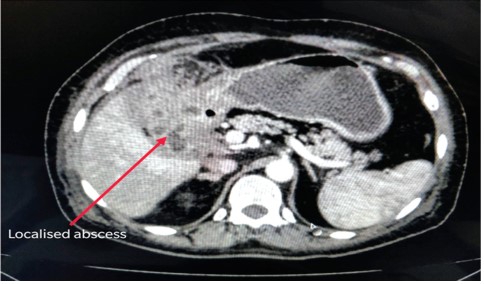
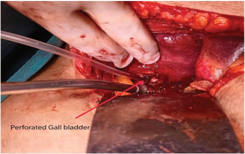
Perforation of the gallbladder is a rare but dreaded complication following cholecystitis.1 Spontaneous perforation is most commonly seen in the elderly, and in patients suffering from diabetes mellitus.2 The most common site of perforation is the fundus of gall bladder.3 The gall bladder distension and edema associated with acute inflammation leads to venous and arterial obstruction. Subsequently, this leads to ischaemia and necrosis of the gall bladder wall. The fundus is most susceptible to this injury due to maximum distance from the cystic arterial supply. Aggravating factors include atherosclerosis, focal vasospasm and localized vasculitis. If diagnosed and treated late, perforation of gallbladder may be associated with high chances of morbidity and mortality.4-6 Here, we report a case of a 66-year-old male with perforation of gallbladder following acute calculous cholecystitis.
References
- 1. Roslyn J, Busuttil RW. Perforation of the gallbladder: a frequently mismanaged condition. Am J Surg. 1979;137(3):307–312.
- 2. Fagan SP et al. Prognostic factors for the development of gangrenous cholecystitis. Am J Surg. 186:481-485,2003.
- 3. Derici H, Kara C, Bozdag AD, Nazli O, Tansug T, Akca E. Diagnosis and treatment of gallbladder perforation. World J Gastroenterol. 2006;12(48):7832–7836.
- 4. Roslyn JJ, Thompson JE, Darvin H, DenBesten L. Risk factors for gallbladder perforation. Am J Gastroenterol. 1987; 82: 636–640.
- 5. Sood BP, Kalra N, Gupta S, Sidhu R, Gulati M, Khandelwal N, Suri S. Role of sonography in the diagnosis of gallbladder perforation. J Clin Ultrasound. 2002; 30: 270–274.
- 6. Lennon F, Green WE. Perforation of the gallbladder. A review of 32 cases. J R CollSurgEdinb. 1983; 28: 169–173.
- 7. Pedrosa CS, Casanova R, Rodriguez R. CT findings in subacute perforation of the gallbladder: report on 5 cases. Eur J Radiol. 1981;1(2):137–142.
- 8. Sood B, Jain M, Khandelwal N, Singh P, Suri S. MRI of perforated gall bladder. AustralasRadiol. 2002;46(4):438–440.
- 9. Niemeier OW. Acute Free Perforation of the Gall-Bladder. Ann Surg. 1934;99(6):922–924.
- 10. Glenn F, Moore SW. Gangrene and perforation of the wall of the gallbladder. Arch Surg. 1942:44677–44686.
- 11. Tsai MJ, Chen JD, Tiu CM, Chou YH, Hu SC, Chang CY. Can acute cholecystitis with gallbladder perforation be detected preoperatively by computed tomography in ED? Correlation with clinical data and computed tomography features. Am J Emerg Med. 2009;27(5):574–581.
- 12. Solva M, Pamilo M, Paivansaio M, Taavitsainen M, Suramo L. Ultrasonography in acute gallbladder perforation. ActaRadiol. 1988:2941–2944.
- 13. Glenn F, Moore SW. Gangrene and perforation of the wall of the gallbladder. Arch Surg. 1942:44677–44686.
- 14. Kalliafas S, Ziegler DW, Flancbaum L, Choban PS. Acute acalculouscholecystitis: incidence, risk factors, diagnosis, and outcome. Am Surg. 1998;64(5):471–475.
- 15. Chen JJ, Lin HH, Chiu CT, Lin DY. Gallbladder perforation with intrahepatic abscess formation. J Clin Ultrasound. 1990;18(1):43– 45.
- 16. Gore RM, Ghahremani GG, Joseph AE, Nemcek AA Jr., Marn CS, Vogelzang RL. Acquired malposition of the colon and gallbladder in patients with cirrhosis: CT findings and clinical implications. Radiology. 1989;171(3):739–742.
Data Sharing Statement
There are no additional data available. All raw data and code are available upon request.
Funding
This research received no funding.
Author Contributions
All authors contributed significantly to the work and approve its publication.
Ethics Declaration
This article does not involve any human or animal subjects, and therefore does not require ethics approval.
Acknowledgements
We would like to express our gratitude to the patients, their families, and all those who have contributed to this study.
Conflicts of Interest
No conflicts of interest in this work.
About this article

Cite this article
Dipanjan Pradhan, Rohit Basu, Saurav Karamakar/Rare Case of Gall Bladder Perforation Following Acute Calculus Cholecystitis in a Elderly Male: A Case Report/New Indian J Surg. 2022;13(4): 167–170.
Licence:
Attribution-Non-commercial 4.0 International (CC BY-NC 4.0)This license enables reusers to distribute, remix, adapt, and build upon the material in any medium or format for noncommercial purposes only, and only so long as attribution is given to the creator.
| Received | Accepted | Published |
|---|---|---|
| June 16, 2022 | July 18, 2022 | December 25, 2022 |
DOI: https://doi.org/10.21088/nijs.0976.4747.13422.4
Keywords
Gall bladder perforationPericholecystic abscessPerforationSearch for Similar Articles
Similar Articles
- End Stage Renal Disease: AVF for Hemodialysys: Clinical Assessment with/without...
- Neutrophil Lymphocyte Ratio: A Prognostic Indicator of Limb Survival in CLI Pati...
- Role of Pan-Tilt Camera in Tele-interaction During Surgery at the Time of COVID-...
- Early Detection of Common Bile Duct Injury Post Cholecystectomy, What are the Op...
- Role of External Tissue Expansion Wound Closure in the Electrical Burn Wound to...
Article Level Metrics
Last UpdatedFriday 09 October 2026, 04:04:56 (IST)
5943
Accesses
3
636
00
Citations
NA
NA
NA
Download citation
Article Keywords
Keyword Highlighting
Highlight selected keywords in the article text.
Timeline
| Received | June 16, 2022 |
| Accepted | July 18, 2022 |
| Published | December 25, 2022 |
licence
Attribution-Non-commercial 4.0 International (CC BY-NC 4.0)
This license enables reusers to distribute, remix, adapt, and build upon the material in any medium or format for noncommercial purposes only, and only so long as attribution is given to the creator.