Full Text (PDF)
Indian Journal of Forensic Odontology 16(1):p 19-27, January-June 2023. | DOI: https://doi.org/10.21088/ijfo.0974.505X.16123.2
Original Article
Pulp Volume: An Indepth Tool in Age Estimation a Comparative Retrospective Forensic based Cone Beam CT
Sindhuja N, Harsha Vardhan B G, Saraswathi K Gopal
Author Information
Licence:
Attribution-Non-commercial 4.0 International (CC BY-NC 4.0)This license enables reusers to distribute, remix, adapt, and build upon the material in any medium or format for noncommercial purposes only, and only so long as attribution is given to the creator.
Indian Journal of Forensic Odontology 16(1):p 19-27, January-June 2023. | DOI: https://doi.org/10.21088/ijfo.0974.505X.16123.2
How Cite This Article:
Sindhuja N, Harsha Vardhan BG, Saraswathi K Gopal/Pulp Volume: An Indepth Tool in Age Estimation a Comparative Retrospective Forensic based Cone Beam CT/Indian Journal of Forensic Odontology. 2023;16(1):19–27.Timeline
Received : January 31, 2023
Accepted : April 27, 2023
Published : June 30, 2023
Abstract
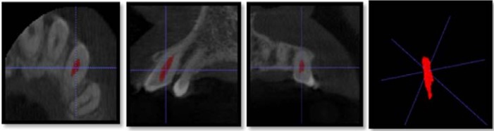
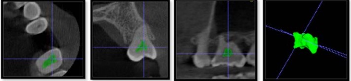
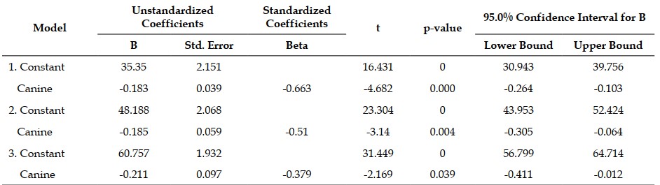
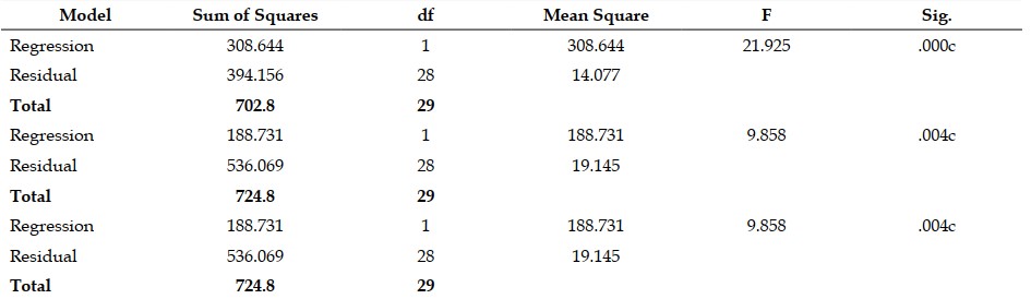
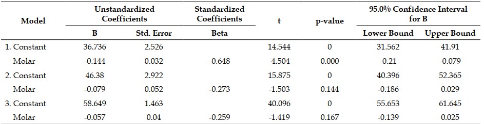
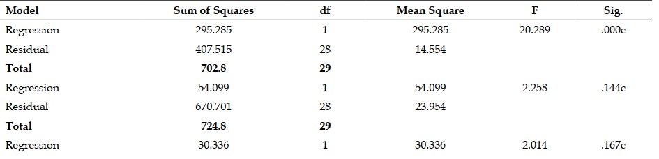
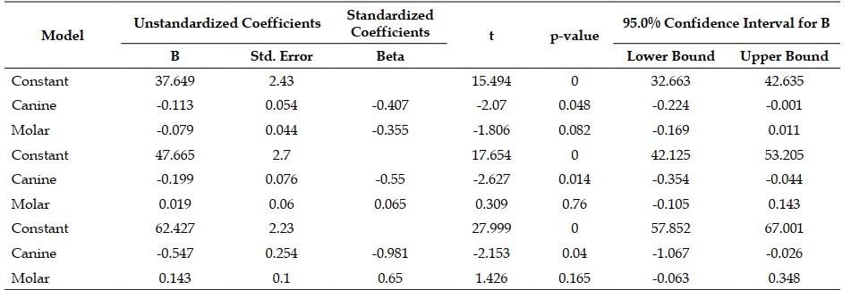
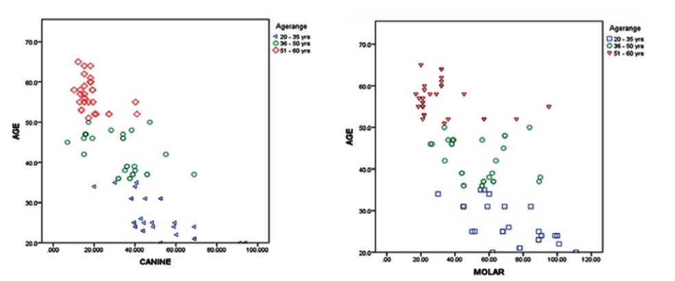
Background: The field of forensics utilizes various craniofacial structures and skull in identifying an unknown deceased victim. This identification deals with assessing the gender and age of skeletonized remains based on eliciting the ethnicity of the population at archaeological sites, and comparison of post-mortem records with the presumed antemortem records. Interestingly, even a single tooth can be used to assess an age of an individual and this is widely used in forensics for investing legal matters as well as in scientific research purpose. Teeth are resistant to environmental insults and post-mortem decomposition and hence can be retained without distortion. Aim and Objectives: To analyse the volumetric data of canine and first molars in cone beam CT for estimating age among samples of various age groups and to compare between those values to evaluate which tooth indexed volumetric data gives more specificity in age estimation. Methodology: It is a retrospective Institutional based Forensic study conducted using 180 samples from 90 full skull CBCT images whose age ranged between 20-65 years, acquired from the dental archives of department of Oral Medicine and Radiology which are equally divided among both genders. Further the samples were categorized into three groups as (20 -35), (36-50) and (51- 65) years in both the gender. All data samples were assessed using the ITK –SNAP 3.8.0 software. Using semi-automatic active contour segmentation method, the volumes of pulps of upper canine and upper first molar were analysed and calculated data were statistically analysed using discriminant functional analysis and multivariate regression analysis to evaluate the correlation of pulp volumes with respect to chronological age. Results: The multivariate regression analysis done for estimating age among the given groups of samples using upper canine gave statistically highly significant p value of 0.001,0.004 and 0.039 for group I (20-35 years), group II (36-50 years) and group III (51-65 years) respectively. The regression analysis done using upper first molar for age estimation gave statistically highly significant p value of 0.001 for group I, whereas group II and group III gave out insignificant results. The comparative analysis between multivariate Regression analysis for age estimation of upper canine and upper first molar to determine which tooth index has more specificity, gave statistically significant results for canine samples in all age groups. The samples were also analysed for Pearson correlation in estimating age, which resulted in high correlation in group I samples for both canine and first molar, moderate correlation for group II and group III samples for canine pulp volumes and low correlation for group II and group III for molar pulp volumes. Conclusion: Over the years, CBCT has evolved a lot and today they are being used in various sectors of dentistry. In the field of forensics, various oral and maxillofacial structures are being used as an adjuvant in personnel identification with more precision using cone beam CT. The results of the present study show statistically highly significant correlation between age and upper canine samples in all the age groups analysed. Hence, based on the results of present study, the upper canine can be used as a promising adjuvant tool in forensic odontology for age estimation.
References
- 1. Shah P, Velani PR, Lakade L, Dukle S. Teeth in forensics: A review. Indian J Dent Res 2019; 30:291- 9.
- 2. Divakar KP. Forensic odontology: The new dimension in dental analysis. International journal of biomedical science: IJBS. 2017 Mar;13(1):1.
- 3. Erbudak HÖ, Ozbek M, Uysal S, Karabulut E (2012) Application of Kvaal et al.’s age estimation method to panoramic radiographs from Turkish individuals. Forensic Sci Int 219: 141-146.
- 4. Lewis JM, Senn DR. Dental age estimation utilizing third molar development: a review of principles, methods, and population studies used in the United States. Forensic science international. 2010 Sep 10;201(1-3):79-83.
- 5. Venkatesh, Elluru; Venkatesh Elluru, Snehal (2017). Cone beam computed tomography: basics and applications in dentistry. Journal of Istanbul University Faculty of Dentistry, 51(0). doi:10.17096/ jiufd.00289
- 6. Divakar KP. Forensic Odontology: The New Dimension in Dental Analysis. Int J Biomed Sci. 2017;13(1):1-5.
- 7. Nagammai N, Saraswathi Gopal K, Srividhya S. Tooth Coronal Pulp Index as A Tool for Age Estimation: An Institutional Based Retrospective Cone Beam Computed Tomography Study. Indian Journal of Forensic Odontology.2019;12(2):35–43.
- 8. Kvaal SI, Kolltveit KM, Thomsen IO, Solheim T (1995) Age estimation of adults from dental radiographs. Forensic Sci Int 74(3): 175–185. https://doi.org/10.1016/0379-0738(95)01760-G.
- 9. Cameriere R, Ferrante L, Cingolani M (2004) Variations in pulp/ tooth area ratio as an indicator of age: a preliminary study. J Forensic Sci 49(2):1–3.
- 10. Shah PH, Venkatesh R. Pulp/tooth ratio of mandibular first and second molars on panoramic radiographs: An aid for forensic age estimation. J Forensic Dent Sci. 2016;8(2):112. doi:10.4103/0975- 1475.186374.
- 11. Biuki N, Razi T, Faramarzi M. Relationship between pulp-tooth volume ratios and chronological age in different anterior teeth on CBCT. J Clin Exp Dent. 2017;9(5): e688-e693. Published 2017 May 1. doi:10.4317/jced.53654.
- 12. Andi Izham; Elza Ibrahim Auerkari; (2021). The use of radiology CBCT in odontology forensic. The 5th biomedical engineering’s recent progress in biomaterials, drugs development, and medical devices: Proceedings of the 5th International Symposium of Biomedical Engineering (ISBE) 2020AIP Conference Proceedings, (), –. doi:10.1063/5.0047278.
- 13. W. De Vos, J. Casselman, G.R. Swennen, Cone-beam computerized tomography (CBCT) imaging of the oral and maxillofacial region: a systematic review of the literature, Int. J. Oral. Maxillofac. Surg. 38 (6) (2009) 609–625.
- 14. Ge, Zhi-pu; Ma, Ruo-han; Li, Gang; Zhang, Ji-zong; Ma, Xu-chen (2015). Age estimation based on pulp chamber volume of first molars from cone-beam computed tomography images. Forensic Science International, 253(), 133.e1–133.e7.
- 15. Kumar, Shiva & Setty, Jyothsna. (2016). Age Estimation Using Pulp Chamber Volume of First Molars from Cone Beam Computed Tomography Images in Indian Population. International Journal of Science and Research (IJSR). 5. 2421-2425.
- 16. Yousefi F, Lari S, Abbas Shokri, Hashemi S, Hosseini M. Age Estimation Based on the Pulp Chamber Volume of Multi-rooted Teeth Using Cone Beam Computed Tomography. Avicenna J Dent Res. 2020;12(1):19-24.
- 17. Tardivo D, Sastre J, Catherine JH, Leonetti G, Adalian P, Foti B. Age determination of adult individuals by three-dimensional modelling of canines. Int J Legal Med. 2014;128(1):161-169. doi:10.1007/s00414-013-0863-2.
- 18. Kazmi, S., Mânica, S., Revie, G. et al. Age estimation using canine pulp volumes in adults: a CBCT image analysis. Int J Legal Med 133, 1967–1976 (2019). https://doi.org/10.1007/s00414-019-02147-5.
- 19. Abdinian M, Katiraei M, Zahedi H, Rengo C, Soltani P, Spagnuolo G. Age Estimation Based on Pulp–Tooth Volume Ratio of Anterior Teeth in Cone-Beam Computed Tomographic Images in a Selected Population: A Cross-Sectional Study. Applied Sciences. 2021; 11(21):9984.
- 20. Star H, Thevissen P, Jacobs R, Fieuws S, Solheim T, Willems G. Human dental age estimation by calculation of pulp-tooth volume ratios yielded on clinically acquired cone beam computed tomography images of monoradicular teeth. J Forensic Sci. 2011;56 Suppl 1:S77-S82.
Data Sharing Statement
There are no additional data available. All raw data and code are available upon request.
Funding
This research received no funding.
Author Contributions
All authors contributed significantly to the work and approve its publication.
Ethics Declaration
This article does not involve any human or animal subjects, and therefore does not require ethics approval.
Acknowledgements
We would like to express our gratitude to the patients, their families, and all those who have contributed to this study.
Conflicts of Interest
No conflicts of interest in this work.
About this article

Cite this article
Sindhuja N, Harsha Vardhan BG, Saraswathi K Gopal/Pulp Volume: An Indepth Tool in Age Estimation a Comparative Retrospective Forensic based Cone Beam CT/Indian Journal of Forensic Odontology. 2023;16(1):19–27.
Licence:
Attribution-Non-commercial 4.0 International (CC BY-NC 4.0)This license enables reusers to distribute, remix, adapt, and build upon the material in any medium or format for noncommercial purposes only, and only so long as attribution is given to the creator.
| Received | Accepted | Published |
|---|---|---|
| January 31, 2023 | April 27, 2023 | June 30, 2023 |
DOI: https://doi.org/10.21088/ijfo.0974.505X.16123.2
Keywords
Pulp VolumeCBCTOral and Maxillofacial RadiologyForensic OdontologyAgeSearch for Similar Articles
Similar Articles
- Determination of Anthropometric Correlation of Golden Proportions Existing in Ma...
- Oral Autopsy in Forensic Odontology as an Effort for Human Identification in Med...
- Exploring the Global Use of Artificial Intelligence in Forensic Investigations
- Comparative Study of Lip Print between Multigender Community using Suzuki Classf...
- Turkey Teeth
Article Level Metrics
Last UpdatedThursday 19 November 2026, 02:05:03 (IST)
2393
Accesses
6
327
00
Citations
NA
NA
NA
Download citation
Article Keywords
Keyword Highlighting
Highlight selected keywords in the article text.
Timeline
| Received | January 31, 2023 |
| Accepted | April 27, 2023 |
| Published | June 30, 2023 |
licence
Attribution-Non-commercial 4.0 International (CC BY-NC 4.0)
This license enables reusers to distribute, remix, adapt, and build upon the material in any medium or format for noncommercial purposes only, and only so long as attribution is given to the creator.