Full Text (PDF)
Pediatric Education and Research 11(3):p 105-108, September – December 2023. | DOI: https://doi.org/10.21088/per.2321.1644.11323.3
Case Report
Neonatal Achondroplasia: Case Report
Hamza Moatasim Solkar, Nilesh Kanase, Abhijit Shinde, Suresh Waydande, Sunil Natha Mhaske
Author Information
Licence:
Attribution-Non-commercial 4.0 International (CC BY-NC 4.0)This license enables reusers to distribute, remix, adapt, and build upon the material in any medium or format for noncommercial purposes only, and only so long as attribution is given to the creator.
Pediatric Education and Research 11(3):p 105-108, September – December 2023. | DOI: https://doi.org/10.21088/per.2321.1644.11323.3
How Cite This Article:
Hamza Moatasim Solkar, Nilesh Kanase, Abhijit shinde, et al. Neonatal Achondroplasia: Case Report. Pediatr. Edu. Res. 2023;11(3): 105-108.Timeline
Received : August 02, 2023
Accepted : October 12, 2023
Published : December 25, 2023
Abstract
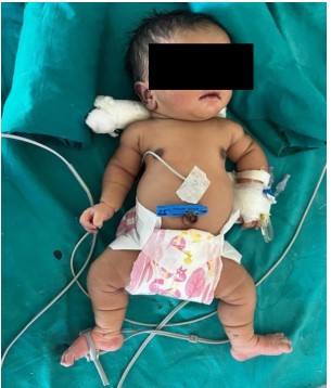
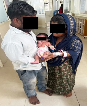
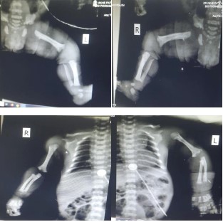
Introduction: Achondroplasia, the most common form of skeletal dysplasia with characteristic short limb dwarfism, is a non lethal variant of chondrodysplasia. Although autosomal dominant genes may be the source of inheritance, most occurrences start off as spontaneous mutations. Case report: Our patient was a term male baby born via elective Caesarean section with birth weight of 2.8kgs. Anomaly scans of 22 weeks of gestation showed no gross anomaly of the fetus.The anomaly scan of 36th week of gestation showed a small size of fetus. On clinical examination, the head circumference was more than normal and the baby had a large head with frontal bossing. There was bilateral symmetrical shortening of upper and lower limbs with short fingers. There was depressed nasal bridge. The abdomen was protuberant and distended. Literature review: A mutation in the 4p16.3 fibroblast growth factor receptor-3 gene (FGFR3) results in achondroplasia. One parent's achondroplasia increases the infant's probability of inheriting the disorder by 50%, and if both parents have it, the infant's chance increases to 75%. This suggests that the disorder may be inherited as an autosomal dominant characteristic.
References
- 1. Cohen MM. Some chondrodysplasias with short limbs: molecular perspectives. American journal of medical genetics. 2002 Oct 15;112(3):304-13.
- 2. Bellus GA, Hefferon TW, de Luna RO, Hecht JT, Horton WA, Machado M, Kaitila I, McIntosh I, Francomano CA. Achondroplasia is defined by recurrent G380R mutations of FGFR3. American journal of human genetics. 1995 Feb;56(2):368.
- 3. Bhusal S, Gautam U, Phuyal R, Choudhary R, Manandhar SR, Niroula A. Diagnosis of achondroplasia at birth: A case report. JNMA: Journal of the Nepal Medical Association. 2020 Feb;58(222):119.
- 4. Horton WA, Hall JG, Hecht JT. Achondroplasia. The Lancet. 2007 Jul 14;370(9582):162-72.
- 5. Baujat G, Legeai-Mallet L, Finidori G, Cormier-Daire V, Le Merrer M. Achondroplasia. Best Practice & Research Clinical Rheumatology. 2008 Mar 1;22(1):3-18.
- 6. Boulet S, Althuser M, Nugues F, Schaal JP, Jouk PS. Prenatal diagnosis of achondroplasia: new specific signs. Prenatal Diagnosis: Published in Affiliation With the International Society for Prenatal Diagnosis. 2009 Jul;29(7):697-702..
- 7. Shelmerdine SC, Brittain H, Arthurs OJ, Calder AD. Achondroplasia: really rhizomelic?. American Journal of Medical Genetics Part A. 2016 Aug;170(8):2039-43.
- 8. Shiang R, Thompson LM, Zhu YZ, Church DM, Fielder TJ, Bocian M, Winokur ST, Wasmuth JJ. Mutations in the transmembrane domain of FGFR3 cause the most common genetic form of dwarfism, achondroplasia. Cell. 1994 Jul 29;78(2):335-42.
- 9. Modaff P, Horton Vk, PaulI RM. Errors in the prenatal diagnosis of children with achondroplasia. Prenatal diagnosis. 1996 Jun;16(6):525-30.
Data Sharing Statement
There are no additional data available. All raw data and code are available upon request.
Funding
This research received no funding.
Author Contributions
All authors contributed significantly to the work and approve its publication.
Ethics Declaration
This article does not involve any human or animal subjects, and therefore does not require ethics approval.
Acknowledgements
We would like to express our gratitude to the patients, their families, and all those who have contributed to this study.
Conflicts of Interest
No conflicts of interest in this work.
About this article

Cite this article
Hamza Moatasim Solkar, Nilesh Kanase, Abhijit shinde, et al. Neonatal Achondroplasia: Case Report. Pediatr. Edu. Res. 2023;11(3): 105-108.
Licence:
Attribution-Non-commercial 4.0 International (CC BY-NC 4.0)This license enables reusers to distribute, remix, adapt, and build upon the material in any medium or format for noncommercial purposes only, and only so long as attribution is given to the creator.
| Received | Accepted | Published |
|---|---|---|
| August 02, 2023 | October 12, 2023 | December 25, 2023 |
DOI: https://doi.org/10.21088/per.2321.1644.11323.3
Keywords
AchondroplasiaShort statureDwarfCaesarean sectionMutationSearch for Similar Articles
Similar Articles
- Mass Abdomen in an Infant: A Pandora Box
- Penetrating Injury Over the Back Due to Wooden Stick – Don’t Leave the Fragments...
- Role of Autologous Platelet Rich Plasma in Management of Pediatric Postburn Web...
- To Study Profile of Hemoglobin and Red Blood Cell Indices during First Week of L...
- Impact of Educational Intervention on Knowledge Regarding Household Waste Manage...
Article Level Metrics
Last UpdatedMonday 07 September 2026, 17:21:37 (IST)
1921
Accesses
4
229
00
Citations
NA
NA
NA
Download citation
Article Keywords
Keyword Highlighting
Highlight selected keywords in the article text.
Timeline
| Received | August 02, 2023 |
| Accepted | October 12, 2023 |
| Published | December 25, 2023 |
licence
Attribution-Non-commercial 4.0 International (CC BY-NC 4.0)
This license enables reusers to distribute, remix, adapt, and build upon the material in any medium or format for noncommercial purposes only, and only so long as attribution is given to the creator.