Full Text (PDF)
Journal of Microbiology and Related Research 12(1):p 17-22, Jan. June 2026. | DOI: 10.21088/jmrr.2395.6623.12126.2
Original Article
Microbial Profile of Pyogenic Infection Causing Isolates from Hospitals in Raipur
Dhananjay Tandon, Pooja Kumari Sahu, Rupal Purena, Madhavi Tiwari, Khushbu Verma, Ashis Kumar Sarkar, Misha Martin
Author Information
Licence:
Attribution-Non-commercial 4.0 International (CC BY-NC 4.0)This license enables reusers to distribute, remix, adapt, and build upon the material in any medium or format for noncommercial purposes only, and only so long as attribution is given to the creator.
Journal of Microbiology and Related Research 12(1):p 17-22, Jan. June 2026. | DOI: 10.21088/jmrr.2395.6623.12126.2
How Cite This Article:
Pooja Kumari Sahu, Dhananjay Tandon, Rupal Purena et. al, Microbial Profile of Pyogenic Infection Causing Isolates from Hospitals in Raipur. J Microbiol Relat Res. 2026; 12(1): 17–22Timeline
Received : June 07, 2026
Accepted : July 10, 2026
Published : June 30, 2026
Abstract
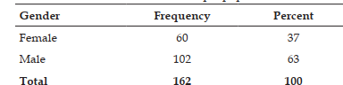
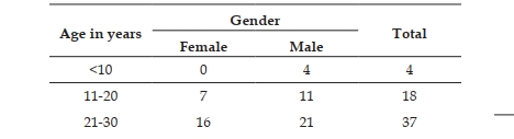
Introduction: Pyogenic infections are caused by pyogenic bacteria, characterized by the formation of pus. These infections can affect various body parts, including the skin, soft tissues, eyes, burns, bones, and joints. Aim: This study aims to isolate and identify bacterial pathogens from pyogenic human infections and analyze the demographics (age and gender) most affected. Methods: Pus sample was collected from 162 patients received from various clinics. The samples were immediately inoculated onto MacConkey agar and blood agar plates and incubated at 37° C for 24 hours. After incubation, Gram staining and biochemical tests were performed to characterize the bacterial isolates. Results: Among the 162 patients, 102 were male (63%) and 60 were female (37%). Out of the 162 pus samples processed, 95 (58.6%) were culture-positive, while 67 (41.4%) showed no bacterial growth. The distribution of 95 culture-positive isolates included: Gram-negative Klebsiella pneumoniae (6), Escherichia coli (35), Acinetobacter spp. (4), and Pseudomonas aeruginosa (8). Gram-positives were Staphylococcus aureus (25), Streptococcus spp. (6), Enterococcus spp. (6). Fungal isolates were candida spp. (2). This study highlights a significant prevalence of pyogenic infections, with a higher incidence among males aged 21– 60 years and females aged 21–50 years. The most frequently isolated pathogens were and, emphasizing the need for effective infection control measures and targeted antibiotic therapy.
References
- 1. Kalita JM, Nag VL, Kombade S, Yedale K. Multidrug resistant superbugs in pyogenic infections: a study from Western Rajasthan, India. The Pan African Medical Journal. 2021;38:409.
- 2. Singh S, Khare M, Patidar RK, Bagde S, Sahare KN, Dwivedi D, Singh V, Center MF, Van Parisar BP. Antibacterial activities against pyogenic pathogens. Int J Pharm Sci Res. 2013;4(8):2974-9.
- 3. Puca V, Marulli RZ, Grande R, Vitale I, Niro A, Molinaro G, Prezioso S, Muraro R, Di Giovanni P. Microbial species isolated from infected wounds and antimicrobial resistance analysis: Data emerging from a three-years retrospective study. Antibiotics. 2021;10(10):1162.
- 4. Yadav L, Ansari S, Yadav SR, Yadav RL. Bacteriological profile of pyogenic infections at a tertiary care centre of Nepal. Microbes and Infectious Diseases. 2022;3(2):332-8.
- 5. Lozano R, Naghavi M, Foreman K, Lim S, Shibuya K, Aboyans V, Abraham J, Adair T, Aggarwal R, Ahn SY, AlMazroa MA. Global and regional mortality from 235 causes of death for 20 age groups in 1990 and 2010: a systematic analysis for the Global Burden of Disease Study 2010. The lancet. 2012;380(9859):2095-128.
- 6. Laxminarayan R, Chaudhury RR. Antibiotic resistance in India: drivers and opportunities for action. PLoS medicine. 2016;13(3):e1001974.
- 7. Sharma V, Parihar G, Sharma V, Sharma H. A study of various isolates from pus sample with their antibiogram from Jln hospital, Ajmer. IOSR Journal of Dental and Medical Sciences. 2015;14(10):64-8.
- 8. Muluye D, Wondimeneh Y, Ferede G, Nega T, Adane K, Biadgo B, Tesfa H, Moges F. Bacterial isolates and their antibiotic susceptibility patterns among patients with pus and/or wound discharge at Gondar university hospital. BMC research notes. 2014;7(1):619.
- 9. Rajput K, Johri S, Goyal A. Identification of bacterial isolates and their antimicrobial susceptibility pattern from wound/pus sample in a tertiary care hospital, Gwalior, Ind Annals of the National Academy of Medical Sciences (India). 2023;59(02):097-103.
- 10. Manjunath NS, Dayanand A, Jagannath KV, Basavarajappa KG, Rangaswamy BE. Prevalence of human pyogenic skin infections, bacterial pathogens and their antibiotic susceptibility profile. J Pub Health Med Res. 2013;1(1):37-42.
- 11. Khelkar Tuneer, K. T., & Tiwari Madhavi, T. M. (2015). A comparative study of hygienic status of butchers and identify bacteria among the slaughters of meat, chicken and fish markets of Jagdalpur city, Chhattisgarh, India.
- 12. AL-Joda BM, Jasim AH. Biochemical testing revision for identification several kinds of bacteria. Journal of University of Babylon for Pure and Applied Sciences. 2021;29(2):168-76.
- 13. Rameshkannan S, Nileshraj G, Rameshprabu S, Mangaiarkkarasi A, Meher Ali R. Pattern of pathogens and their sensitivity isolated from pus culture reports in a tertiary care hospital, puducherry. Indian J Basic Appl Med Res. 2014;4(1):243-8.
- 14. Zhang S, Ren L, Li Y, Wang J, Yu W, Li N, Li J. Bacteriology and drug susceptibility analysis of pus from patients with severe intra-abdominal infection induced by abdominal trauma. Experimental and therapeutic medicine. 2014;7(5):1427-31.
- 15. Rijal BP, Satyal D, Parajuli NP. High burden of antimicrobial resistance among Bacteria causing pyogenic wound infections at a tertiary Care Hospital in Kathmandu, Nepal. Journal of pathogens. 2017;2017(1):9458218.
Data Sharing Statement
There are no additional data available. All raw data and code are available upon request.
Funding
This research received no funding.
Author Contributions
All authors contributed significantly to the work and approve its publication.
Ethics Declaration
This article does not involve any human or animal subjects and therefore does not require ethics approval.
Acknowledgements
We would like to express our gratitude to the patients, their families, and all those who have contributed to this study.
Conflicts of Interest
No conflicts of interest in this work.
About this article

Cite this article
Pooja Kumari Sahu, Dhananjay Tandon, Rupal Purena et. al, Microbial Profile of Pyogenic Infection Causing Isolates from Hospitals in Raipur. J Microbiol Relat Res. 2026; 12(1): 17–22
Licence:
Attribution-Non-commercial 4.0 International (CC BY-NC 4.0)This license enables reusers to distribute, remix, adapt, and build upon the material in any medium or format for noncommercial purposes only, and only so long as attribution is given to the creator.
| Received | Accepted | Published |
|---|---|---|
| June 07, 2026 | July 10, 2026 | June 30, 2026 |
DOI: 10.21088/jmrr.2395.6623.12126.2
Keywords
• Pyogenic Infection •Pus SampleAge and Gender DemographySearch for Similar Articles
Similar Articles
- Vitiligo: A Comprehensive Review
- Seaweed-Derived Bioplastics: A Sustainable Alternative to Petroleum Based Plasti...
- Antibiogram of Bacterial Isolates from Critical Care Patients in Sharda Hospital...
- Prevalence and Antimicrobial Resistance Patterns of Staphylococcus aureus Isolat...
- Bacteriology of Burn Wound Infections and their Antibiogram
Article Level Metrics
Last UpdatedSaturday 17 October 2026, 08:39:50 (IST)
1831
Accesses
16
223
00
Citations
NA
NA
NA
Download citation
Article Keywords
Keyword Highlighting
Highlight selected keywords in the article text.
Timeline
| Received | June 07, 2026 |
| Accepted | July 10, 2026 |
| Published | June 30, 2026 |
licence
Attribution-Non-commercial 4.0 International (CC BY-NC 4.0)
This license enables reusers to distribute, remix, adapt, and build upon the material in any medium or format for noncommercial purposes only, and only so long as attribution is given to the creator.