Full Text (PDF)
Journal of Microbiology and Related Research 12(1):p 7-16, Jan. June 2026. | DOI: 10.21088/jmrr.2395.6623.12126.1
Original Article
A Study on the Isolation and Identification of Biofilm forming Oral Flora
Neha Chauhan, Pallavi Sharma, Shweta , Gupta, Ashifa Gouri
Author Information
Licence:
Attribution-Non-commercial 4.0 International (CC BY-NC 4.0)This license enables reusers to distribute, remix, adapt, and build upon the material in any medium or format for noncommercial purposes only, and only so long as attribution is given to the creator.
Journal of Microbiology and Related Research 12(1):p 7-16, Jan. June 2026. | DOI: 10.21088/jmrr.2395.6623.12126.1
How Cite This Article:
Neha Chauhan, Pallavi Sharma, Shweta Gupta et. al, A Study on the Isolation and Identification of Biofilm forming Oral Flora. J Surg. Nurs. 2026; 12(1): 07-16.Timeline
Received : April 13, 2026
Accepted : May 17, 2026
Published : June 30, 2026
Abstract
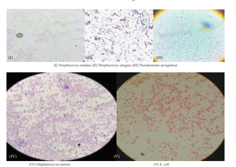
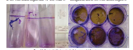
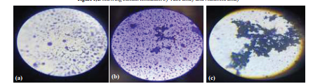
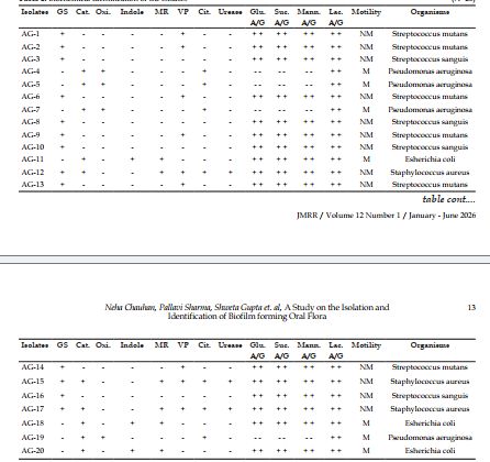
Biofilm is a community of micro-organisms, such as bacteria, that adheres to surfaces and form a protective slimy matrix. Biofilms are the most complex structure of micro-organisms attached to the animate and inanimate objects such as- teeth, pipes, medical devices etc. These cells are frequently embedded within a self-produced extracellular polymeric substance (EPS). This extracellular polymeric substance protects the bacterial cells from environmental conditions. Physical properties of biofilms such as attachment, mechanical strength, and antibiotic resistance can be attributed to EPS matrix. Dental biofilm forming organisms are major sources of the oral diseases like disease of the gums that is initiated by subsets of microbes that colonize on or just below the gingival margin. Dental Biofilms are the most prevalent cause of oral diseases that colonizes in the gingival and sub-gingival regions of the mouth. The present study aims to isolate the biofilm forming bacteria from the oral cavity, their morphological characterization, biochemical identification and detection of biofilm by various assays. Firstly, samples were collected from the oral cavity of healthy individuals, and then inoculated on various media such as- NA, TSA, and MS-Agar for isolation of biofilm forming microorganisms. Ten isolates were Gram positive cocci either in chains or clumps. Seven isolates were Gram negative rods and three isolates were Gram positive cocci in bunches. The isolates were characterized by Gram staining, Biochemical tests involving IMViC test, catalase test, oxidase, urease, sugar fermentation and motility test. The isolated organisms were observed for their ability to lyse the RBCs by haemolysis test. Biofilm formation assay was carried out by tube assay, tissue culture plate assay, coverslip assay and by microtiter dish assay. It was observed that out of 20 samples 14 samples were found to produce biofilm. Four out of fourteen isolates were strong and six isolates were moderate biofilm producers (10) by tube assay. Five out of fourteen isolates were strong and six were moderate biofilm producers (11) by tissue culture plate assay and microtiter dish assay. While only very few samples showed the same formation by coverslip assay. Therefore, the TC plate assay and Microtiter dish assay can be used upon the tube method for the detection of biofilm formation.
References
- 1. Huang, R., Li, M. and Gregory, R.L., 2011. Bacterial interactions in dental biofilm. Virulence, 2(5), pp.435-444.
- 2. Marsh PD. Dental plaque: biological significance of a biofilm and community lifestyle. J ClinPeriodontol. 2005;32(Suppl 6):7–15.
- 3. Saini, R., Saini, S. and Sharma, S., 2011. Biofilm: A dental microbial infection. Journal of natural science, biology, and medicine, 2(1), p.71.
- 4. Bassler BL, Losick R. Bacterially speaking. Cell. 2006;125(2):237–46.
- 5. Hall-Stoodley L, Costerton JW, Stoodley P. Bacterial biofilms: from the natural environment to infectious diseases. Nat Rev Microbiol. 2004;2(2):95–108.
- 6. Davey M.E., O’Toole G.A. Microbial biofilms: From ecology to molecular genetics. Microbiol. Mol. Biol. Rev. 2000;64:847–867. doi: 10.1128/ MMBR.64.4.847-867.2000. [PMC free article] [PubMed] [CrossRef] [Google Scholar].
- 7. Berlanga M., Guerrero R. Living together in biofilms: The microbial cell factory and its biotechnological implications. Microbe.Cell Factories. 2016;15:165. doi: 10.1186/s12934-15 JMRR / Volume 12 Number 1 / January - June 2026 016-0569-5. [PMC free article] [PubMed] [CrossRef] [Google Scholar].
- 8. Flemming H.-C., Wingender J. The biofilm matrix. Nat. Rev. Microbiol. 2010;8:623– 633. doi: 10.1038/nrmicro2415. [PubMed] [CrossRef] [Google Scholar].
- 9. Kaplan J.B. Biofilm dispersal: Mechanisms, clinical implications, and potential therapeutic uses. J. Dent. Res. 2010;89:205–218. doi: 10.1177/0022034509359403. [PMC free article] [PubMed] [CrossRef] [Google Scholar].
- 10. Stewart P.S., Franklin M.J. Physiological heterogeneity in biofilms. Nat. Rev. Microbiol. 2008;6:199–210. doi: 10.1038/nrmicro1838. [PubMed] [CrossRef] [Google Scholar].
- 11. Boles B.R., Singh P.K. Endogenous oxidative stress produces diversity and adaptability in biofilm communities. Proc. Natl. Acad. Sci. USA. 2008;105:12503–12508. doi: 10.1073/ pnas.0801499105. [PMC free article] [PubMed] [CrossRef] [Google Scholar].
- 12. Costerton J.W., Cheng K.J., Geesey G.G., Ladd T.I., Nickel J.C., Dasgupta M., Marrie T.J. Bacterial biofilms in nature and disease. Annu. Rev. Microbiol. 1987;41:435–464. doi: 10.1146/ annurev.mi.41.100187.002251. [PubMed] [CrossRef] [Google Scholar].
- 13. Stewart P.S., William Costerton J. Antibiotic resistance of bacteria in biofilms. Lancet. 2001;358:135–138. doi: 10.1016/S0140- 6736(01)05321-1. [PubMed] [CrossRef] [Google Scholar]
- 14. Hall C.W., Mah T.-F. Molecular mechanisms of biofilm-based antibiotic resistance and tolerance in pathogenic bacteria.FEMS Microbiol. Rev. 2017;41:276–301. doi: 10.1093/ femsre/fux010. [PubMed] [CrossRef] [Google Scholar]
- 15. Garrett T., Bhakoo M., Zhang Z. Bacterial adhesion and biofilms on surfaces. Prog. Nat. Sci. 2008;18doi: 10.1016/j.pnsc.2008.04.001. [CrossRef] [Google Scholar]
- 16. Conley J., Olson M.E., Cook L.S., Ceri H., Phan V., Davies H.D. Biofilm Formation by Group A Streptococci: Is There a Relationship with Treatment Failure? J. Clin. Microbiol. 2003;41:4043–4048. doi: 10.1128/ JCM.41.9.4043-4048.2003. [PMC free article] [PubMed] [CrossRef] [Google Scholar]
- 17. Engel, A.S., Kranz, H.T., Schneider, M., Tietze, J.P., Piwowarcyk, A., Kuzius, T., Arnold, W. and Naumova, E.A., 2020. Biofilm formation on different dental restorative materials in the oral cavity.BMC Oral Health, 20(1), pp.1-10.
- 18. Sonkusale, K. and Tale, V. (2015). Isolation and Characterization of Biofilm Forming Bacteria from Oral Microflora. Int. J. Curr. Microbiol. App. Sci Special Issue-2: 118-127118.
- 19. Krzyściak, W., Jurczak, A. and Piątkowski, J., 2016. The role of human oral microbiome in dental biofilm formation.Microbial Biofilms— Importance and Applications. In Tech, pp.329-382.
- 20. Who, (1997). Oral health surveys. Basic method. 4th ed. Geneva.
- 21. Sullivan, A.; Borgstrom, M.; Granath, L. and Nilsson, G. (1996). Number of mutans streptococci or lactobacilli in a total dental plaque sample does not explain the variation in caries better than stimulated whole saliva. Comm. Dent. Oral. /Epid. 24: 159-163.
- 22. Krzyściak, W., Jurczak, A., Kościelniak, D., Bystrowska, B. and Skalniak, A., 2014. The virulence of Streptococcus mutans and the ability to form biofilms.European Journal of Clinical Microbiology & Infectious Diseases, 33, pp.499-515.
- 23. Li YH, et al. Cell density modulates acid adaptation in Streptococcus mutans: implications for survival in biofilms. J Bacteriol. 2001;183(23):6875–84.
- 24. Burne RA, Chen YY, Penders JE. Analysis of gene expression in Streptococcus mutans in biofilms in vitro. Adv Dent Res. 1997;11(1):100–9.
- 25. Mohr M, et al. Quantification of cell fusion events human breast cancer cells and breast epithelial cells using a Cre-LoxP-based double fluorescence reporter system. Cell Mol Life Sci. 2015;72(19):3769–82. https://doi.org/10.1007/ s00018-015-1910-6. Epub 2015 Apr 22.
- 26. Matsumoto-Nakano M. Role of Streptococcus mutans surface proteins for biofilm formation. Jpn. Dent. Sci. Rev. 2018;54:22–29. doi: 10.1016/j.jdsr.2017.08.002. [PMC free article] [PubMed] [CrossRef] [Google Scholar]
- 27. Christensen, G.D., Simpson, W.A., Bisno, A.L. and Beachey, E.H., 1982. Adherence of slime-producing strains of Staphylococcus epidermidis to smooth surfaces.Infection and immunity, 37(1), pp.318-326. 28. Chauhan, N., Khan, M.A. and Farooq, U., 2016. Biofilm: An important factor in the prevalence of MDR Salmonella typhi. Int J Biol Pharm Allied Sci, pp.2464-73. Neha Chauhan, Pallavi Sharma, Shweta Gupta et. al, A Study on the Isolation and Identification of Biofilm forming Oral Flora16 Journal of Microbiology and Related Research JMRR / Volume 12 Number 1 / January - June 2026
- 29. Taj, Y., Essa, F., Aziz, F. and Kazmi, S.U., 2012. Study on biofilm-forming properties of clinical isolates of Staphylococcus aureus. The Journal of Infection in Developing Countries, 6(05), pp.403-409.
- 30. O’Toole, G.A., 2011. Microtiter dish biofilm formation assay. JoVE (Journal of Visualized Experiments), (47), p.e2437.
- 31. Burmolle M, Thomsen TR, Fazli M, Dige I, Christensen L, Homoe P, et al., Biofilms in chronic infections- a matter of opportunitymonospeciesbiofilms in multispecies infections, FEMS Immunol Med Microbiol, 59, 2010, 324-36
Data Sharing Statement
There are no additional data available. All raw data and code are available upon request.
Funding
This research received no funding.
Author Contributions
All authors contributed significantly to the work and approve its publication.
Ethics Declaration
This article does not involve any human or animal subjects, and therefore does not require ethics approval.
Acknowledgements
We would like to express our gratitude to the patients, their families, and all those who have contributed to this study.
Conflicts of Interest
The authors report no conflicts of interest in this work.
About this article

Cite this article
Neha Chauhan, Pallavi Sharma, Shweta Gupta et. al, A Study on the Isolation and Identification of Biofilm forming Oral Flora. J Surg. Nurs. 2026; 12(1): 07-16.
Licence:
Attribution-Non-commercial 4.0 International (CC BY-NC 4.0)This license enables reusers to distribute, remix, adapt, and build upon the material in any medium or format for noncommercial purposes only, and only so long as attribution is given to the creator.
| Received | Accepted | Published |
|---|---|---|
| April 13, 2026 | May 17, 2026 | June 30, 2026 |
DOI: 10.21088/jmrr.2395.6623.12126.1
Keywords
Biofilm FormationDental PlaqueExopolysaccharidesDental FloraTube MethodAdhesion AssayCoverslip AssaySearch for Similar Articles
Similar Articles
- Vitiligo: A Comprehensive Review
- Seaweed-Derived Bioplastics: A Sustainable Alternative to Petroleum Based Plasti...
- Antibiogram of Bacterial Isolates from Critical Care Patients in Sharda Hospital...
- Prevalence and Antimicrobial Resistance Patterns of Staphylococcus aureus Isolat...
- Bacteriology of Burn Wound Infections and their Antibiogram
Article Level Metrics
Last UpdatedSunday 18 October 2026, 11:58:31 (IST)
1838
Accesses
28
223
00
Citations
NA
NA
NA
Download citation
Article Keywords
Keyword Highlighting
Highlight selected keywords in the article text.
Timeline
| Received | April 13, 2026 |
| Accepted | May 17, 2026 |
| Published | June 30, 2026 |
licence
Attribution-Non-commercial 4.0 International (CC BY-NC 4.0)
This license enables reusers to distribute, remix, adapt, and build upon the material in any medium or format for noncommercial purposes only, and only so long as attribution is given to the creator.