Full Text (PDF)
Indian Journal of Diabetes and Endocrinology 8(1):p 23-29, Jan. June 2026. | DOI: n.a.
Original Article
Impact of One Mineral Density among Postmenopausal Women with Gestational DM using Dual Energy X-ray Absorptiometry
Manjunath Jadhav, Ashok Kumar, Veena Kardiguddi, Vidyashri Kattimani, Dada Hayat Sakali
Author Information
Licence:
Attribution-Non-commercial 4.0 International (CC BY-NC 4.0)This license enables reusers to distribute, remix, adapt, and build upon the material in any medium or format for noncommercial purposes only, and only so long as attribution is given to the creator.
Indian Journal of Diabetes and Endocrinology 8(1):p 23-29, Jan. June 2026. | DOI: n.a.
How Cite This Article:
Ashok Kamat, Manjunath Jadhav, Veena Kardiguddi et. al, Impact of bone mineral density among postmenopausal women with gestational DM using dual energy X-ray absorptiometry. Ind Jl of Diabetes and Endo. 2026;8(1): 23–29Timeline
Received : April 24, 2026
Accepted : May 27, 2026
Published : June 25, 2026
Abstract
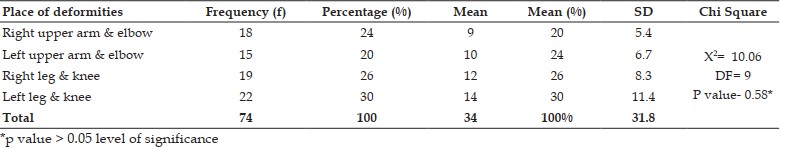
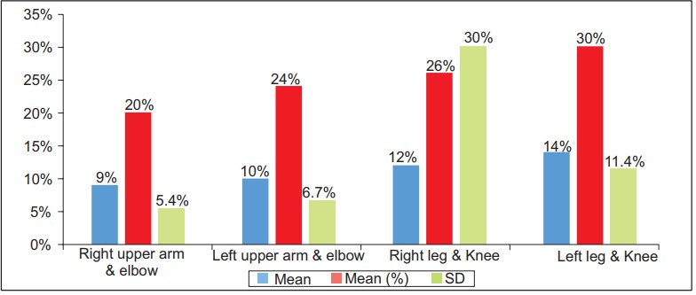
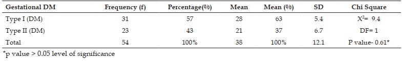
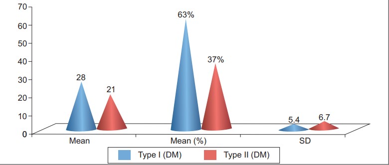
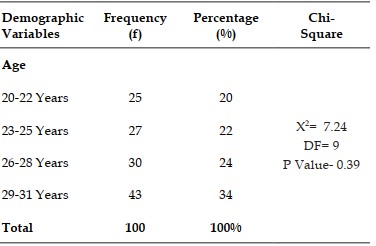
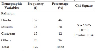
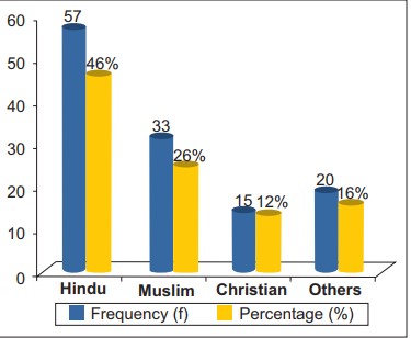
Bone mineral density has been assessed using Dual-Energy X-Ray Absorptiometry. Bone mineral density is measured according to the results of the Dual-Energy X-Ray Absorptiometry examination of the vertebral column and pelvis. Although gestational DM is known to affect bone mineral density, at the present time this particular relationship is not clear, in this study evaluating the effects of gestational diabetes mellitus on bone mineral density among postmenopausal women’s. Bone density of controls and patients was measured using a DEXA scan in this study the number of samples were 125, the investigator employed purposive sampling technique for data collection demographic Performa and semi-structure questionnaire was used, and for data analysis inferential statistics used from SPSS software. Aim: Is to evaluate the impact of bone mineral density among postmenopausal women with gestational DM using X-ray absorptiometry and to determine association between impact of bone mineral density among postmenopausal women with gestational DM using X-ray absorptiometry and demographic variables. Materials and Methods: This study was conducted in the Outpatient Clinic of Rheumatology, Arihant Multispecialty tertiary care teaching, Belagavi. From February 2026 to April as a part of evaluating the effects of gestational diabetes mellitus on bone mineral density among postmenopausal women’s, the number of samples were 125, the investigator employed purposive sampling technique for data collection demographic Performa and semi-structure questionnaire was used, and for data analysis inferential statistics used from SPSS software. Results: Graph 1, revealed that majority of the respondents having left leg and knee deformity their mean is 14 (30%) and SD will be 11.4, more or less similar results showing right leg and knee deformity their mean is 12 (26%) and SD will be 8.3, , more or less similar results showing left upper arm and elbow deformity their mean is 10 (24%) and SD will be 6.7, and less similar results showing right upper arm and elbow deformity their mean is 9 (20%) and SD will be 5.4. Conclusion: At the end of investigation the researcher concluded that based on bone mineral density of the upper extremities and lower extremities with osteoporosis among females majority of the finding are 14 (30%) are having left leg and knee deformity, and based on Type of DM among postmenopausal women’s with their mean, mean (%), and SD, revealed that majority of the respondents having Type I (DM) is about 28 (63%) and SD will be 5.4, and 21 (37%) and SD will be 6.7.
References
- 1. Wei W, Shary JR, Garrett-Mayer E, Anderson B, Forestieri NE, Hollis BW, Wagner CL. Bone mineral density during pregnancy in women participating in a randomized controlled trial of vitamin D supplementation. The American Journal of Clinical Nutrition. 2017 December 1; 106(6):1422-30.
- 2. To WW, Wong MW. Bone mineral density changes during pregnancy in actively exercising women as measured by quantitative ultrasound. Archives of gynaecology and obstetrics. 2012 August; 286(2):357-63.
- 3. Keyhanmanesh R, Daghigh F, Rasihashemi SZ. Contradictory results of bone mineral density values and bone turnover markers in post-menopausal diabetic women: A literature review. Journal of Clinical Sciences. 2024 October 1; 21(4):190-6.
- 4. Al-Maatouq MA, El-Desouki MI, Othman SA, Mattar EH, Babay ZA, Addar M. Prevalence of osteoporosis among postmenopausal females with diabetes mellitus. Saudi Medical Journal. 2004; 25(10):1423-7.
- 5. Watts NB, Binkley N, Owens CD, Al-Hendy A, Puscheck EE, Shebley M, Schlaff WD, Simon JA. Bone mineral density changes associated with pregnancy, lactation, and medical treatments in premenopausal women and effects later in life. Journal of women’s health. 2021 October; 30(10):1416-30.
- 6. Engberg E, Koivusalo SB, Huvinen E, Viljakainen H. Bone health in women with a history of gestational diabetes or obesity. Acta Obstetricia et Gynecologica Scandinavica. 2020 April; 99(4):477-87.
- 7. Eroglu S, Karatas G, Aziz V, Gursoy AF, Ozel S, Gulerman HC. Evaluation of bone mineral density and its associated factors in postpartum women. Taiwanese Journal of Obstetrics and Gynaecology. 2019 Nov 1; 58(6):801-4
- 8. Aziz ZA, Dawood NS, Al-Khalisy MH. Evaluation of the effect of type II diabetes mellitus on bone mineral density of upper and lower limbs by dual-energy X-ray absorptiometry. Journal of the Faculty of Medicine Baghdad. 2023 Apr 27; 65(1):27-33.
- 9. Laitinen K, Välimäki M, Keto P. Bone mineral density measured by dual-energy X-ray absorptiometry in healthy Finnish women. Calcified tNumber international. 1991 April; 48(4):224-31.
- 10. Alhowiti NM, Mackawy AM, Al Wanian WM, Alshebremi M, Allemailem KS, Alharbi HO. Type 2 Diabetes Mellitus and Osteoporosis: Site-Specific Bone Mineral Density Variations and Metabolic Correlations in Postmenopausal Saudi Women. Medicina. 2025 April 24; 61(5):789.
Data Sharing Statement
There are no additional data available. All raw data and code are available upon request.
Funding
This research received no funding.
Author Contributions
All authors contributed significantly to the work and approve its publication.
Ethics Declaration
Provide information related to the Ethics Committee approval with approval number OR write This article does not involve any human or animal subjects, and therefore does not require ethics approval.
Acknowledgements
We would like to express our gratitude to the patients, their families, and all those who have contributed to this study.
Conflicts of Interest
The authors report no conflicts of interest in this work.
About this article
Cite this article
Ashok Kamat, Manjunath Jadhav, Veena Kardiguddi et. al, Impact of bone mineral density among postmenopausal women with gestational DM using dual energy X-ray absorptiometry. Ind Jl of Diabetes and Endo. 2026;8(1): 23–29
Licence:
Attribution-Non-commercial 4.0 International (CC BY-NC 4.0)This license enables reusers to distribute, remix, adapt, and build upon the material in any medium or format for noncommercial purposes only, and only so long as attribution is given to the creator.
| Received | Accepted | Published |
|---|---|---|
| April 24, 2026 | May 27, 2026 | June 25, 2026 |
DOI: n.a.
Keywords
Bone Mineral DensityPostmenopausal WomenGestational DmDualEnergy X-Ray and AbsorptiometrySearch for Similar Articles
Similar Articles
- The Gut-Glycemic Axis in the Indian Geriatric Population: Evaluating Pre and Pro...
- Evaluate the Knowledge about Hyperparathyroidism and Hyperthyroidism among Nursi...
- Comparative Study to Evaluate the Knowledge Level about Acromegaly and Gigantis...
- HbA1c in Diabetes: Indian Perspectives on Measurement, Interpretation, and Clini...
- Evaluate the Knowledge about Type 1 DM and Type 2 DM among Staff Nurses Working...
Article Level Metrics
Last UpdatedTuesday 28 July 2026, 09:09:33 (IST)
793
Accesses
24
128
00
Citations
NA
NA
NA
Download citation
Article Keywords
Keyword Highlighting
Highlight selected keywords in the article text.
Timeline
| Received | April 24, 2026 |
| Accepted | May 27, 2026 |
| Published | June 25, 2026 |
licence
Attribution-Non-commercial 4.0 International (CC BY-NC 4.0)
This license enables reusers to distribute, remix, adapt, and build upon the material in any medium or format for noncommercial purposes only, and only so long as attribution is given to the creator.