Full Text (PDF)
Journal of Cardiovascular Medicine and Surgery 8(4):p 117-122, October-December 2022. | DOI: https://doi.org/10.21088/jcms.2454.7123.8422.2
Original Article
Guidelines Based Management of Venous Ulcer
Ravi Kumar Chittoria, Barath Kumar Singh. P
Author Information
Licence:
Attribution-Non-commercial 4.0 International (CC BY-NC 4.0)This license enables reusers to distribute, remix, adapt, and build upon the material in any medium or format for noncommercial purposes only, and only so long as attribution is given to the creator.
Journal of Cardiovascular Medicine and Surgery 8(4):p 117-122, October-December 2022. | DOI: https://doi.org/10.21088/jcms.2454.7123.8422.2
How Cite This Article:
Barath Kumar Singh. P, Ravi Kumar Chittoria/Guidelines Based Management of Venous Ulcer/J Cardiovasc Med Surg. 2022;8(4): 117-122.Timeline
Received : September 23, 2022
Accepted : October 25, 2022
Published : December 30, 2022
Abstract
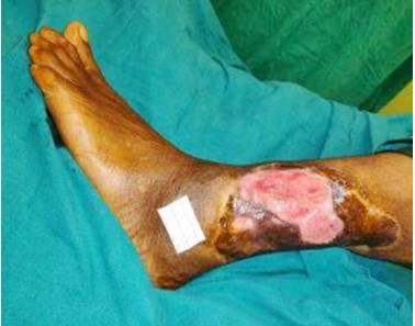
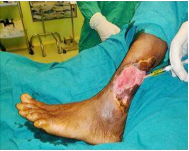
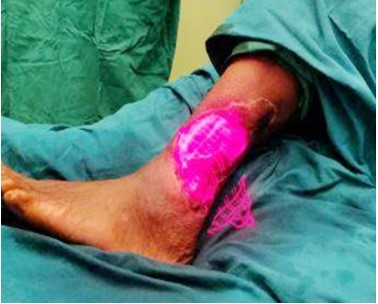
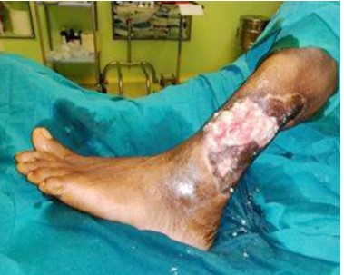
Venous disorders causing a permanent increase in venous pressure are by far the most frequent reason for ulcers of lower extremity. Venous ulceration is the most severe and debilitating outcome of chronic venous insufficiency in the lower limbs and accounts for 80 percent of lower extremity ulcerations. The recurrence of venous ulcers is a common thing due to the recurrence of varicosities and non-compliance of the patient to the treatment. Various other causes for lower extremity ulcerations could be arterial insufficiency, prolonged pressure, diabetic neuropathy and systemic illness such as rheumatoid arthritis, vasculitis, osteomyelitis, and skin malignancy. A thorough investigation of underlying venous disorder is a prerequisite for a differentiated therapy. Venous ulcer causes a significant burden for the health care system. This article highlights the role of SWCR guidelines in management of venous ulcer.
References
- 1. Briggs M, Nelson EA. Topical agents or dressings for pain in venous leg ulcers. Cochrane Database Syst Rev. 2003;1:CD001177.
- 2. Nelzén O, Bergqvist D, Lindhagen A. Long-term prognosis for patients with chronic leg ulcers: a prospective cohort study. Eur J VascEndovasc Surg. 1997;13(5):500-508.
- 3. Samson RH, Showalter DP. Stockings and the prevention of recurrent venous ulcers. Dermatol Surg. 1996;22(4):373-376.
- 4. Abbade LP, Lastória S. Venous ulcer: epidemiology, physiopathology, diagnosis and treatment. Int J Dermatol. 2005;44(6):449-456.
- 5. Callam MJ, Ruckley CV, Harper DR, Dale JJ. Chronic ulceration of the leg: extent of the problem and provision of care. Br Med J (Clin Res Ed). 1985;290(6485):1855-1856.
- 6. Ruckley CV. Socioeconomic impact of chronic venous insufficiency and leg ulcers. Angiology. 1997;48(1):67-69.
- 7. Avci P, Gupta A, Sadasivam M, Vecchio D, Pam Z, Pam N, Hamblin MR. Low-level laser (light) therapy (LLLT) in skin: stimulating, healing, restoring. In Seminars in cutaneous medicine and surgery 2013 Mar (Vol. 32, No. 1, p. 41). NIH Public Access.
- 8. Winfeld B. Topical oxygen and hyperbaric oxygen therapy use and healing rates in diabetic foot ulcers. Wounds. 2014;26:E39–47.
- 9. Flanagan M. The physiology of wound healing. J Wound Care. 2000;9:299–300.
- 10. Schreml S, Szeimies RM, Prantl L, Karrer S, Landthaler M, Babilas P. Oxygen in acute and chronic wound healing. Br J Dermatol. 2010;163:257– 68.
- 11. Hunt SD, Elg F. Clinical effectiveness of hemoglobin spray (Granulox®) as adjunctive therapy in the treatment of chronic diabetic foot ulcers. Diabet Foot Ankle. 2016;7:33101. Published 2016 Nov 7. doi:10.3402/dfa.v7.33101.
- 12. Lubart R, Wollman Y, Friedmann H, Rochkind S, Laulicht I. Effects of visible and near infrared lasers on cell cultures. J PhotochemPhotobiol B. 1992;12(3):305–310.
- 13. Wu W, Naim JO, Lanzafame RJ. The effect of laser irrafiation on the release of bFGF from 3T3 fibroblasts. PhotochemPhotobiol. 1994;59(2):167– 170.
- 14. Vinck EM, Cagnie BJ, Cornelissen MJ, Declercq HA, Cambier DC. Increased fibroblast proliferation induced by light emitting diode and low power laser irradiation. Lasers Med Sci. 2003;18(2):95–99.
- 15. Frigo L, Fávero GM, Lima HJ, Maria DA, Bjordal JM, et al. Low-level laser irradiation (InGaAlP-660 nm) increases fibroblast cell proliferation and reduces cell death in a dose-dependent manner. Photomed Laser Surg. 2010;28(Suppl 1):S151–S156.
- 16. Basso FG, Oliveira CF, Kurachi C, Hebling J, Costa CA. Biostimulatory effect of low-level laser therapy on keratinocytes in vitro. Lasers Med Sci. 2013;28(2):367–374.
- 17. Szymanska J, Goralczyk K, Klawe JJ, Lukowicz M, Michalska M, et al. Phototherapy with low-level laser influences the proliferation of endothelial cells and vascular endothelial growth factor and transforming growth factor-beta secretion. J PhysiolPharmacol. 2013;64(3):387–391.
- 18. Moore P, Ridgway TD, Higbee RG, Howard EW, Lucroy MD. Effect of wavelength on low-intensity laser irradiation-stimulated cell proliferation in vitro. Lasers Surg Med. 2005;36(1):8–12.
- 19. Agaiby AD, Ghali LR, Wilson R, Dyson M. Laser modulation of angiogenic factor production by T-lymphocytes. Lasers Surg Med. 2000;26(4):357– 363.
- 20. Siadat AH, Isseroff RR. Prolotherapy: Potential for the Treatment of Chronic Wounds? Adv Wound Care (New Rochelle). 2019 Apr 1;8(4):160-167. doi: 10.1089/wound.2018.0866. Epub 2019 Apr 3.
- 21. Tumino G, Masuelli L, Bei R, Simonelli L, Santoro A, Francipane S. Topical treatment of chronic venous ulcers with sucralfate: a placebo controlled randomized study. Int J Mol Med. 2008 Jul;22(1):17- 23.
- 22. Sureshbabu A, Okajima H, Yamanaka D, Shastri S, Tonner E, Rae C, et al. IGFBP-5 induces epithelial and fibroblast responses consistent with the fibrotic response. BiochemSoc Trans 2009;37:882-5.
- 23. Van Etten, E., & Mathieu, C. (2005). Immunoregulation by 1,25-dihydroxyvitamin D3: Basic concepts. The Journal of Steroid Biochemistry and Molecular Biology, 97, 93–101.
- 24. Helming, L., Böse, J., Ehrchen, J., Schiebe, S., Frahm, T., & Geffers, R. (2005). 1α, 25- dihydroxyvitamin D3 is a potent suppressor of interferon γ-mediated macrophage activation. Blood, 106, 4351–4358.
- 25. Pittas, A. G., Lau, J., Hu, F. B., & Dawson-Hughes, B. (2007). The role of vitamin D and calcium in type 2 diabetes. A systematic review and metaanalysis. The Journal of Clinical Endocrinology and Metabolism, 92, 2017–2029.
- 26. Schutz GS, Sibbald RG, Falanga V, et al. Wound bed preparation: a systemic approach to wound management. Wound Repair Regen. 2003;11(Suppl 1):S1-28.
- 27. Granick MS, Tran BNN, Alvarez OM. Latest advances in wound debridement techniques. Surg Technol Int. 2020;36:37-40.
- 28. Li Z, Guo S, Yao F, Zhang Y, Li T. Increased ratio of serum matrix metalloproteinase-9 against TIMP-1 predicts poor wound healing in diabetic foot ulcers. J Diabetes Complications. 2013;27(4):380-2.
- 29. Yang C, Zhu P, Yan L, Chen L, Meng R, Lao G. Dynamic changes in matrix metalloproteinase 9 and tissue inhibitor of metalloproteinase 1 levels during wound healing in diabetic rats. J Am Podiatr Med Assoc. 2009;99(6):489-96.
Data Sharing Statement
There are no additional data available. All raw data and code are available upon request.
Funding
This research received no funding.
Author Contributions
All authors contributed significantly to the work and approve its publication.
Ethics Declaration
This article does not involve any human or animal subjects, and therefore does not require ethics approval.
Acknowledgements
We would like to express our gratitude to the patients, their families, and all those who have contributed to this study.
Conflicts of Interest
No conflicts of interest in this work.
About this article

Cite this article
Barath Kumar Singh. P, Ravi Kumar Chittoria/Guidelines Based Management of Venous Ulcer/J Cardiovasc Med Surg. 2022;8(4): 117-122.
Licence:
Attribution-Non-commercial 4.0 International (CC BY-NC 4.0)This license enables reusers to distribute, remix, adapt, and build upon the material in any medium or format for noncommercial purposes only, and only so long as attribution is given to the creator.
| Received | Accepted | Published |
|---|---|---|
| September 23, 2022 | October 25, 2022 | December 30, 2022 |
DOI: https://doi.org/10.21088/jcms.2454.7123.8422.2
Keywords
VenousUlcerGuidelinesSearch for Similar Articles
Similar Articles
- Cardioembolic Stroke as an Initial Presentation of Carney's Complex: A Case Repo...
- Simultaneous Atrioventricular Valve Repair Compelling Creation of Unconventional...
- Iatrogenic Right Radial Arteriovenous Fistula Post Percutaneous Coronary Interve...
- Revascularization in Chronic Coronary Disease: A Comparison between AHA/ACC (202...
- Improvement in Qualitative Indices with Novel Therapy of Nicorandil in Patients...
Article Level Metrics
Last UpdatedMonday 28 September 2026, 16:17:48 (IST)
1823
Accesses
7
242
00
Citations
NA
NA
NA
Download citation
Article Keywords
Keyword Highlighting
Highlight selected keywords in the article text.
Timeline
| Received | September 23, 2022 |
| Accepted | October 25, 2022 |
| Published | December 30, 2022 |
licence
Attribution-Non-commercial 4.0 International (CC BY-NC 4.0)
This license enables reusers to distribute, remix, adapt, and build upon the material in any medium or format for noncommercial purposes only, and only so long as attribution is given to the creator.