Full Text (PDF)
Journal of Cardiovascular Medicine and Surgery 10(1-2):p 39-42, January – June 2024. | DOI: https://doi.org/10.21088/jcms.2454.7123.10(1-2)24.7
Case Report
Simultaneous Atrioventricular Valve Repair Compelling Creation of Unconventional Fenestration During Extracardiac Total Cavopulmonary Connection
Gananjay G. Salve, Shreepal A. Jain, Bharat V. Dalvi, Krishnanaik Shivaprakash
Author Information
Licence:
Attribution-Non-commercial 4.0 International (CC BY-NC 4.0)This license enables reusers to distribute, remix, adapt, and build upon the material in any medium or format for noncommercial purposes only, and only so long as attribution is given to the creator.
Journal of Cardiovascular Medicine and Surgery 10(1-2):p 39-42, January – June 2024. | DOI: https://doi.org/10.21088/jcms.2454.7123.10(1-2)24.7
How Cite This Article:
Gananjay G. Salve, Shreepal A. Jain, Bharat V. Dalvi, Krishnanaik Shivaprakash. Simultaneous Atrioventricular Valve Repair Compelling Creation of Unconventional Fenestration During Extracardiac Total Cavopulmonary Connection. J Cardiovasc Med Surg.2024;10(1-2):39-42.Timeline
Received : February 26, 2024
Accepted : June 25, 2024
Published : June 30, 2024
Abstract
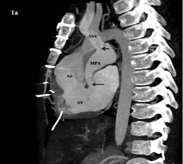
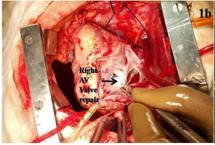
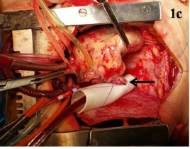
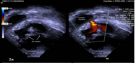
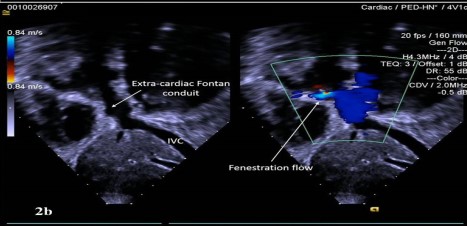
Severe regurgitation of the atrio-ventricular valve and borderline pulmonary vascular resistance are risk factors for failure of univentricular repair. We believe that fenestration in the Fontan pathway plays an important role in avoiding postoperative catastrophesimmediate and late, in such high-risk substrates. Rarely, the intra-operative circumstances compel the surgeon to opt for an unconventional method of fenestration. We report an interesting case of high-risk univentricular repair, had previously undergone pulmonary artery banding and bidirectional Glenn shunt, who now required a simultaneous repair of the regurgitant atrio-ventricular valve compelling the creation of an unconventional fenestration during Stage III palliation.
References
- 1. Tseng SY, Siddiqui S, Di Maria MV, Hill GD, Lubert AM, Kutty S, Opotowsky AR, Possner M, Morales DLS, Quintessenza JA, Alsaied T. Atrioventricular Valve Regurgitation in Single Ventricle Heart Disease: A Common Problem Associated With Progressive Deterioration and Mortality. J Am Heart Assoc. 2020 Jun 2;9(11):e015737. doi: 10.1161/ JAHA.119.015737.
- 2. Podzolkov VP, Chiaureli MR, Yurlov IA, Zelenikin MM, Kovalev DV, Dontsova VI, Astrakhantseva TO, Putiato NA, Zaets SB. Results of Fontan operation in patients with atrioventricular valve regurgitation. Eur J Cardiothorac Surg. 2015;48:308- 14; discussion 314-5.
- 3. Alsaied T, Bokma JP, Engel ME, Kuijpers JM, Hanke SP, Zuhlke L, Zhang B, Veldtman GR. Factors associated with long-term mortality after Fontan procedures: a systematic review. Heart. 2017;103:104–110.
- 4. d’Udekem Y, Xu MY, Galati JC, Lu S, Iyengar AJ, Konstantinov IE, Wheaton GR, Ramsay JM, Grigg LE, Millar J, et al. Predictors of survival after singleventricle palliation: the impact of right ventricular dominance. J Am Coll Cardiol. 2012;59:1178–1185.
- 5. King G, Ayer J, Celermajer D, Zentner D, Justo R, Disney P, Zannino D, d’Udekem Y. Atrioventricular valve failure in Fontan palliation. J Am Coll Cardiol. 2019;73:810–822.
- 6. Li D, Li M, Zhou X, An Q. Comparison of the fenestrated and non-fenestrated Fontan procedures: A meta-analysis. Medicine (Baltimore). 2019;98:e16554.
- 7. Marcelletti CF, Iorio FS, Abella RF. Late results of extracardiac Fontan repair. Semin Thorac Cardiovasc Surg Pediatr Card Surg Annu 1999;2:131–42.
- 8. Kreutzer C, Schlichter AJ, Simon JL, Conejeros Parodi WM, Blunda C, Kreutzer GO. A new method for reliable fenestration in extracardiac conduit Fontan operations. Ann Thorac Surg 2003;75:1675–9.
- 9. Salve GG, Jain SA, Adnaik A, Shivaprakash K. An unusual fenestration in single-stage Fontan operation. Interact Cardiovasc Thorac Surg 2016;23:656-8.
Data Sharing Statement
There are no additional data available. All raw data and code are available upon request.
Funding
This research received no funding.
Author Contributions
All authors contributed significantly to the work and approve its publication.
Ethics Declaration
This article does not involve any human or animal subjects, and therefore does not require ethics approval.
Acknowledgements
We would like to express our gratitude to the patients, their families, and all those who have contributed to this study.
Conflicts of Interest
No conflicts of interest in this work.
About this article

Cite this article
Gananjay G. Salve, Shreepal A. Jain, Bharat V. Dalvi, Krishnanaik Shivaprakash. Simultaneous Atrioventricular Valve Repair Compelling Creation of Unconventional Fenestration During Extracardiac Total Cavopulmonary Connection. J Cardiovasc Med Surg.2024;10(1-2):39-42.
Licence:
Attribution-Non-commercial 4.0 International (CC BY-NC 4.0)This license enables reusers to distribute, remix, adapt, and build upon the material in any medium or format for noncommercial purposes only, and only so long as attribution is given to the creator.
| Received | Accepted | Published |
|---|---|---|
| February 26, 2024 | June 25, 2024 | June 30, 2024 |
DOI: https://doi.org/10.21088/jcms.2454.7123.10(1-2)24.7
Keywords
Atrio-ventricular valve regurgitationCompletion fontan procedureUnconventional fenestrationSearch for Similar Articles
Similar Articles
- Cardioembolic Stroke as an Initial Presentation of Carney's Complex: A Case Repo...
- Iatrogenic Right Radial Arteriovenous Fistula Post Percutaneous Coronary Interve...
- Revascularization in Chronic Coronary Disease: A Comparison between AHA/ACC (202...
- Improvement in Qualitative Indices with Novel Therapy of Nicorandil in Patients...
- Management of Tetralogy of Fallot with Major Aortopulmonary Collateral Arteries:...
Article Level Metrics
Last UpdatedSunday 18 October 2026, 19:24:30 (IST)
2267
Accesses
9
263
00
Citations
NA
NA
NA
Download citation
Article Keywords
Keyword Highlighting
Highlight selected keywords in the article text.
Timeline
| Received | February 26, 2024 |
| Accepted | June 25, 2024 |
| Published | June 30, 2024 |
licence
Attribution-Non-commercial 4.0 International (CC BY-NC 4.0)
This license enables reusers to distribute, remix, adapt, and build upon the material in any medium or format for noncommercial purposes only, and only so long as attribution is given to the creator.