Full Text (PDF)
Journal of Cardiovascular Medicine and Surgery 12(1):p 31-33, Jan-April 2026. | DOI: 10.21088/jcms.2454.7123.12126.5
Case Report
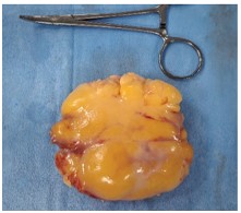
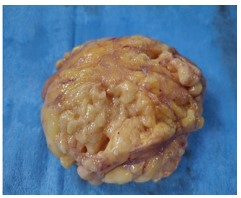
Giant Intra-cardiac Lipoma in the Right Atrium
A.P. Sharath, Raghavendra Murthy, Chandana N.C, Prabhavathi Bhat, Sattenapalli Sravya
Author Information
Licence:
Attribution-Non-commercial 4.0 International (CC BY-NC 4.0)This license enables reusers to distribute, remix, adapt, and build upon the material in any medium or format for noncommercial purposes only, and only so long as attribution is given to the creator.
Journal of Cardiovascular Medicine and Surgery 12(1):p 31-33, Jan-April 2026. | DOI: 10.21088/jcms.2454.7123.12126.5
How Cite This Article:
Sharath AP, Murthy R, Chandana NC, et al. Giant Intra-cardiac Lipoma in the Right Atrium. Indian J Cardiovasc Med Surg. 2026;12(1):31–33.Timeline
Received : February 13, 2026
Accepted : March 16, 2026
Published : April 30, 2026
Abstract
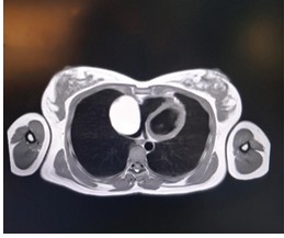
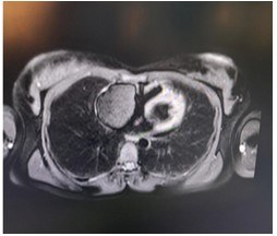
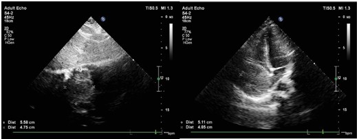
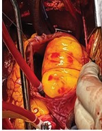
Lipomas are rare tumours of the heart constituting a small percentage of the primary cardiac tumours of the heart. They comprise of mature adipocytes in an encapsulated form when benign; the malignant version being the liposarcoma. Lipoma is the second most common primary benign cardiac neoplasm and presents most commonly in middle-aged and older adults. It has no sex predilection. Approximately 50% of lipomas originate from the subendocardial layer, while the other half arise from the subepicardial or myocardial layers and grow into the pericardial sac. Patients are mostly asymptomatic but may sometimes experience arrhythmias or valvular dysfunction, with varying symptoms depending on the tumor’s location. Subepicardial lipomas can occasionally compress the coronary arteries, leading to ischemic chest pain. Non-invasive cardiac imaging can help in early detection and accurate diagnosis of cardiac lipomas, which is crucial for management to avoid unfavorable outcomes due to overgrowth. We encountered a giant lipoma in the right atrium of a 35 year old school teacher.
References
- 1. Centofanti P., Rosa E.D., Deorsola L., Dato GMA, Patene F., Torre M., Barbato L., and et al. Primary cardiac tumors: early and late results of surgical treatment in 91 patients. The Annals of Thoracic Surgery, 1999, 68(4); 1236-41.
- 2. Shu S., Wang J., Zheng C. From pathogenesis to treatment, a systemic review of cardiac lipoma. Journal of Cardiothoracic Surgery. 2021; 16(1): 2-7
- 3. Rajiah P., Kanne J.P., Kalahasti V., Schoenhagen P. Computed tomography of cardiac and pericardiac masses. J Cardiovasc Comput Tomogr 2011; 5: 16–29.
- 4. Ossola P., Pannone L., Spoladore R., et al. The silent cardiac mass. J Cardiovasc Med (Hagerstown). 2019; 20(10): 718–20.
- 5. Tyebally S., Chen D., Bhattacharyya S., Mughrabi A., Hussain Z., Manisty C. and et al. Cardiac Tumors: State-of-the-Art Review. JACC Cardiooncology. 2020; 2(2): 293-311.
- 6. Steger C.M. Intrapericardial giant lipoma displacing the heart. Int Sch Res Notices 2011; 2011: e243637.
Data Sharing Statement
There are no additional data available. All raw data and code are available upon request.
Funding
This research received no funding.
Author Contributions
All authors contributed significantly to the work and approve its publication.
Ethics Declaration
This article does not involve any human or animal subjects, and therefore does not require ethics approval.
Acknowledgements
We would like to express our gratitude to the patients, their families, and all those who have contributed to this study.
Conflicts of Interest
The authors report no conflicts of interest in this work.
About this article
Cite this article
Sharath AP, Murthy R, Chandana NC, et al. Giant Intra-cardiac Lipoma in the Right Atrium. Indian J Cardiovasc Med Surg. 2026;12(1):31–33.
Licence:
Attribution-Non-commercial 4.0 International (CC BY-NC 4.0)This license enables reusers to distribute, remix, adapt, and build upon the material in any medium or format for noncommercial purposes only, and only so long as attribution is given to the creator.
| Received | Accepted | Published |
|---|---|---|
| February 13, 2026 | March 16, 2026 | April 30, 2026 |
DOI: 10.21088/jcms.2454.7123.12126.5
Keywords
Lipoma• Intracardiac massIntracardiac massPrimary benign cardiac tumoursSearch for Similar Articles
Similar Articles
- Case Report: Neurophysiological Facilitation of Respiration in an Infant with...
- Left Coronary Artery Fistula Masquerading as Coronary Insufficiency: A Rare Sur...
- Hemodynamic Monitoring by Point-of-Care Ultrasound: A Multiorgan, Physiology-Dri...
- Aortotomy Closure after Cardiac Valve Replacements: Nightmare for Surgeons
- Rare Neck Angiolipoma Managed with Surgical Excision: A Case Report
Article Level Metrics
Last UpdatedMonday 27 July 2026, 17:16:17 (IST)
963
Accesses
12
136
00
Citations
NA
NA
NA
Download citation
Article Keywords
Keyword Highlighting
Highlight selected keywords in the article text.
Timeline
| Received | February 13, 2026 |
| Accepted | March 16, 2026 |
| Published | April 30, 2026 |
licence
Attribution-Non-commercial 4.0 International (CC BY-NC 4.0)
This license enables reusers to distribute, remix, adapt, and build upon the material in any medium or format for noncommercial purposes only, and only so long as attribution is given to the creator.