Full Text (PDF)
Indian Journal of Forensic Odontology 16(1):p 9-17, January-June 2023. | DOI: https://doi.org/10.21088/ijfo.0974.505X.16123.1
Original Article
Frontal Sinus & Nasal Septum Patterns & Pneumatisation of Sphenoid Sinus: An Obscure Path for Personal Identification: A Retrospective Cone Beam Computed Tomography Study
Saraswathi Gopal. K null, Kalai Selvi Ravishankar, Mahesh Kumar P null
Author Information
Licence:
Attribution-Non-commercial 4.0 International (CC BY-NC 4.0)This license enables reusers to distribute, remix, adapt, and build upon the material in any medium or format for noncommercial purposes only, and only so long as attribution is given to the creator.
Indian Journal of Forensic Odontology 16(1):p 9-17, January-June 2023. | DOI: https://doi.org/10.21088/ijfo.0974.505X.16123.1
How Cite This Article:
Ravishankar KS, Gopal KS, Kumar PM. Frontal Sinus & Nasal Septum Patterns & Pneumatisation of Sphenoid Sinus: An Obscure Path for Personal Identification: A Retrospective Cone Beam Computed Tomography Study. Indian J Forensic Odontol. 2023;16(1):9–17.Timeline
Received : May 29, 2023
Accepted : June 26, 2023
Published : June 30, 2023
Abstract
Background: Identification of a body is the cornerstone of forensic investigation. If primary methods of identification are impossible (absence of comparative DNA samples, fingerprints, and teeth records), it can be performed using radiographic techniques, whenever antemortem images are available. Visual comparison of antemortem and postmortem images, such as frontal sinuses radiographics and CBCTs can allow identification. The uniqueness and reliability of frontal sinus and nasal septum patterns is used for human identification in forensic science. personal identification can also be done by assessment of pneumatisation of sphenoidal sinus.
Aim & Objectives: This study aims to evaluate the use of frontal sinus and nasal septum patterns and the pneumatisation of sphenoidal sinus for personal identification. To correlate all the parameters to find which is more accurate for identifying a person.
Materials & Method: Study Design: A Retrospective Study. Study Population: A study of 90 CBCT images, (45 males and 45 females) acquired from the archives that were generated using Planmeca Promax 3D MID Proface CBCT machine and assessed with Planmeca Romexis Software. Sample size: Sample of 90 CBCT images obtained from the archives. CBCT images of patients between 18 - 69 years of age.
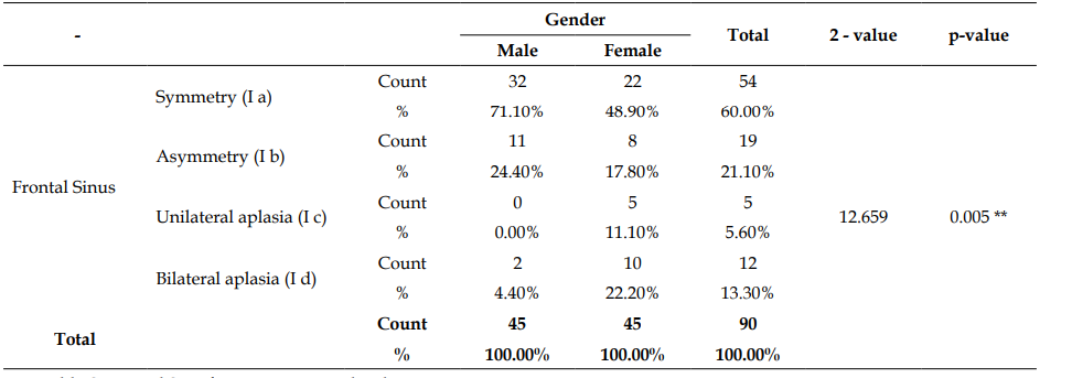
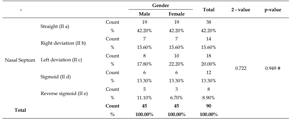
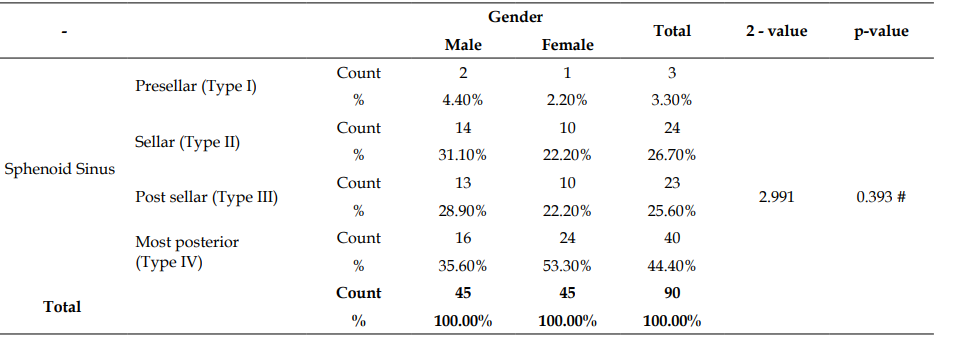
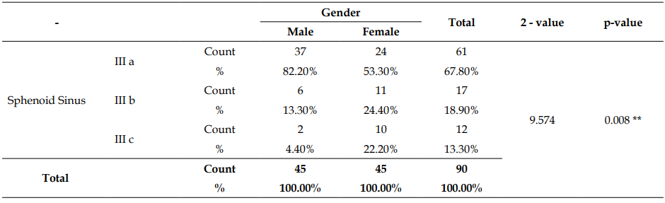
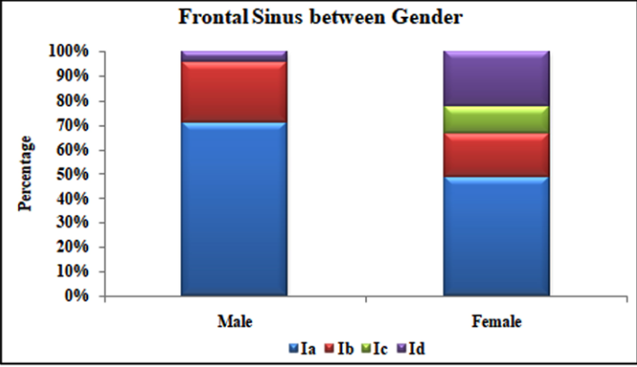
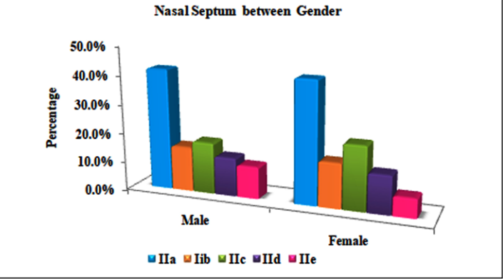
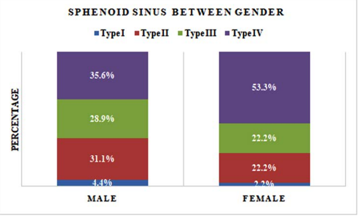
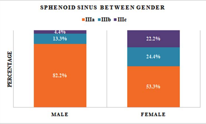
Result: Pearson’s Chi-Square test between gender for frontal sinus were 2=12.659, p=0.005<0.01 which shows highly statistical significance between frontal sinus and gender, for nasal septum were 2=0.722, p=0.949>0.05 which shows no statistical significance between nasal septum and gender, for sphenoid sinus were 2=9.574, p=0.008<0.01 which shows highly statistical significance between sphenoid sinus and gender. But his study had individual variations in frontal sinus, nasal septum and sphenoid sinus for every person.
References
- 1. Pramod JB, Marya AV, Sharma V. Role of forensic odontologist in post mortem person identification. Dent Res J. 2013;9(5):522-530.
- 2. Sidhu R, Chandra S, Devi P, Taneja N, Sah K, Kaur N. Forensic importance of maxillary sinus in gender determination: a morphometric analysis from Western Uttar Pradesh, India. Eur J Gen Dent. 2014;3(1):53-56.
- 3. David MP, Saxena R. Use of frontal sinus and nasal septum patterns as an aid in personal identification: a digital radiographic pilot study. J Forensic Dent Sci. 2010;2(2):77-80.
- 4. Interpol. Disaster Victim Identification guide [Internet]. 2014 [cited 2024].
- 5. Brown KA. Dental identification of unknown bodies. Ann Acad Med Singap. 1984;13(1):3-7.
- 6. Lorkiewicz-Muszyńska D, Łabecka M, Żaba C, et al. Difficulties in identification of human corpses and skeletal remains on the basis of dental records and examinations. Arch Med Sadowej Kryminol. 2009;59(3):218-224.
- 7. Thali MJ, Markwalder T, Jackowski C, Sonnenschein M, Dirnhofer R. Dental CT imaging as a screening tool for dental profiling: advantages and limitations. J Forensic Sci. 2006;51(1):113-119.
- 8. Pfaeffli M, Vock P, Dirnhofer R, et al. Post-mortem radiological CT identification based on classical ante-mortem X-ray examinations. Forensic Sci Int. 2007;171(2-3):111-117.
- 9. Kanthem RK, Guttikonda VR, Yeluri S, Kumari G. Sex determination using maxillary sinus. J Forensic Dent Sci. 2015;7(2):163-167.
- 10. Pasquier E, De Saint Martin Pernot L, Burdin V, et al. Determination of age at death: assessment of an algorithm of age prediction using numerical three-dimensional CT data from pubic bones. Am J Phys Anthropol. 1999;108(3):261-268.
- 11. Mehta M, Saini V, Nath S, Menon SK. CT scan images for sex discrimination—a preliminary study on Gujarati population. J Forensic Radiol Imaging. 2015;3(1):43-48.
- 12. Stoković N, Trkulja V, Dumić-Čule I, et al. Sphenoid sinus types, dimensions and relationship with surrounding structures. Ann Anat. 2016;203:69-76.
- 13. Vidya CS, Shamsundar NM. Computerized tomographic study of frontal sinus patterns in skulls of south Indian population. Int J Sci Res Publ. 2014;4(9):1-5.
- 14. Belaldavar C, Kotrashetti VS, Hallikerimath SR, Kale AD. Assessment of frontal sinus dimensions to determine sexual dimorphism among Indian adults. J Forensic Dent Sci. 2014;6(1):25-30.
- 15. Taniguchi M, Sakoda S, Kano T, et al. Possible use of nasal septum and frontal sinus patterns to radiographic identification of unknown human remains. Osaka City Med J. 2003;49(1):31-38.
- 16. Dawidson I. Case reports and background: difficulties with identification—Sweden. J Forensic Odontostomatol. 2011;29(1):44-50.
- 17. Byard RW, Gilbert JD, Kostakis C, Heath KJ. Circumstances of death and diagnostic difficulties in brushfire fatalities. J Forensic Sci. 2012;57(4):969-972.
- 18. Barghouth G, Prior JO, Lepori D, et al. Paranasal sinuses in children: size evaluation of maxillary, sphenoid, and frontal sinuses by magnetic resonance imaging and proposal of volume index percentile curves. Eur Radiol. 2002;12(6):1451-1458.
- 19. Scuderi AJ, Harnsberger HR, Boyer RS. Pneumatization of the paranasal sinuses: normal features of importance to the accurate interpretation of CT scans and MR images. AJR Am J Roentgenol. 1993;160(5):1101-1104.
- 20. Casselman JW. Le sphénoïde: anatomie [The sphenoid: anatomy]. J Neuroradiol. 2003;30(4):201-210.
- 21. Peele JC. Unusual anatomical variations of the sphenoid sinuses. Laryngoscope. 1957;67(3):208-237.
- 22. Kazkayasi M, Karadeniz Y, Arikan OK. Anatomic variations of the sphenoid sinus on computed tomography. Rhinology. 2005;43(2):109-114.
- 23. Sirikci A, Bayazit YA, Bayram M, et al. Variations of sphenoid and related structures. Eur Radiol. 2000;10(5):844-848.
- 24. Güldner C, Pistorius SM, Diogo I, et al. Analysis of pneumatization and neurovascular structures of the sphenoid sinus using cone-beam tomography (CBT). Acta Radiol. 2012;53(2):214-219.
- 25. Hatch GM, Dedouit F, Christensen AM, et al. RADid: a pictorial review of radiologic identification using postmortem CT. J Forensic Radiol Imaging. 2014;2:52-59.
- 26. Modi JP. A textbook of medical jurisprudence and toxicology. 24th ed. Noida: LexisNexis Butterworths; 2011.
- 27. Gopal KS, Vijayan BP. Significance of frontal sinus and nasal septum patterns in personal identification in forensics: a retrospective CBCT study. Int J Oral Health Med Res. 2016;2:57-61.
- 28. Auffret M, et al. Contribution of the computed tomography of the anatomical aspects of the sphenoid sinuses to forensic identification. J Neuroradiol. 2016;43(5):331-337.
Data Sharing Statement
There are no additional data available. All raw data and code are available upon request.
Funding
This research received no funding.
Author Contributions
Whether all authors contributed significantly to the work and approve its publication.
Ethics Declaration
This article does not involve any human or animal subjects, and therefore does not require ethics approval.
Acknowledgements
We would like to express our gratitude to the patients, their families, and all those who have contributed to this study.
Conflicts of Interest
The authors report no conflicts of interest in this work.
About this article

Cite this article
Ravishankar KS, Gopal KS, Kumar PM. Frontal Sinus & Nasal Septum Patterns & Pneumatisation of Sphenoid Sinus: An Obscure Path for Personal Identification: A Retrospective Cone Beam Computed Tomography Study. Indian J Forensic Odontol. 2023;16(1):9–17.
Licence:
Attribution-Non-commercial 4.0 International (CC BY-NC 4.0)This license enables reusers to distribute, remix, adapt, and build upon the material in any medium or format for noncommercial purposes only, and only so long as attribution is given to the creator.
| Received | Accepted | Published |
|---|---|---|
| May 29, 2023 | June 26, 2023 | June 30, 2023 |
DOI: https://doi.org/10.21088/ijfo.0974.505X.16123.1
Keywords
Frontal sinusNasal septumSphenoid sinusPersonal identificationCBCTSearch for Similar Articles
Similar Articles
- Forensics in Orthodontics: A Narrative Review
- Forensic Identification Through CBCT Evaluation of Secondary Dentin, Root Canal...
- Reliability and Accuracy of Palatal Rugoscopy as a Tool for Gender Identificati...
- Training Indian Undergraduate Students in Dental Charting and Radiographic Comp...
- Use of Saliva Biomarkers and DNA Epigenetics in Forensic Identification: A Narr...
Article Level Metrics
Last UpdatedSaturday 17 October 2026, 10:47:47 (IST)
1937
Accesses
13
267
00
Citations
NA
NA
NA
Download citation
Article Keywords
Keyword Highlighting
Highlight selected keywords in the article text.
Timeline
| Received | May 29, 2023 |
| Accepted | June 26, 2023 |
| Published | June 30, 2023 |
licence
Attribution-Non-commercial 4.0 International (CC BY-NC 4.0)
This license enables reusers to distribute, remix, adapt, and build upon the material in any medium or format for noncommercial purposes only, and only so long as attribution is given to the creator.