Full Text (PDF)
Indian Journal of Dental Education 16(2):p 91-94, April – June 2023. | DOI: https://doi.org/10.21088/ijde.0974.6099.16223.6
Case Report
Dental Calculus Induced Peripheral Ossifying Fibroma: A Clinical Case Report
Zubair Ahmad Janbaz, Huda Hussain, Suhail Majid Jan, Roobel Behal
Author Information
Licence:
Attribution-Non-commercial 4.0 International (CC BY-NC 4.0)This license enables reusers to distribute, remix, adapt, and build upon the material in any medium or format for noncommercial purposes only, and only so long as attribution is given to the creator.
Indian Journal of Dental Education 16(2):p 91-94, April – June 2023. | DOI: https://doi.org/10.21088/ijde.0974.6099.16223.6
How Cite This Article:
Janbaz ZA, Hussain H, Jan SM, et al. Dental calculus induced peripheral ossifying fibroma: a clinical case report. Indian J Dent Educ. 2023;16(2):91-94.Timeline
Received : March 14, 2023
Accepted : March 29, 2023
Published : April 30, 2023
Abstract
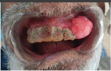
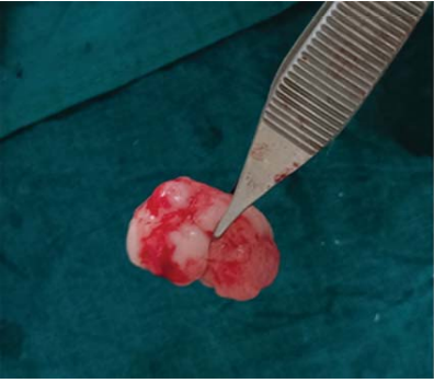
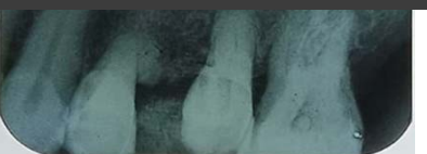
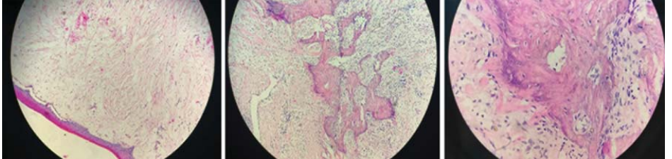
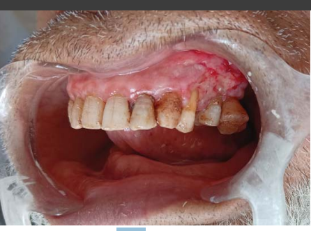
Fibromas are benign growths of fibrous, connective tissue. Gingiva is a common site of oral fibromas. Owing to similar and overlapping presentation of various oral lesions, arriving at a specific diagnosis becomes difficult. Lesions like pyogenic granuloma, irritational fibroma, Peripheral Ossifying Fibroma (POF) and Angio-granuloma are more or less indistinguishable. POF is a relatively rare gingival lesion with multiple histopathologic presentations. The pathogenesis of POF is not amply clear though it is considered reactive in nature. Etiological stimulus may be irritation from dental calculus, plaque, orthodontic appliances or faulty restorations. A clinical report of a 67 year old male with a large peripheral ossifying fibroma in the anterior maxilla showing profuse growth resulting in facial asymmetry and causing masticatory discomfort, is presented. Surgical excision of the lesion was done and subsequent histopathologic confirmation of excised sample confirmed the clinical diagnosis.
References
- 1. Buchner A, Hansen LS. The histomorphologic spectrum of peripheral ossifying fibroma. Oral Surg Oral Med Oral Pathol. 1987;63(4):452-461.
- 2. Neville BW, Damm DD, Allen CM, Bouquot JE. Oral and Maxillofacial Pathology. 2nd ed. Philadelphia: Saunders; 2002. Soft tissue tumors; p. 451-452.
- 3. Yadav R, Gulati A. Peripheral ossifying fibroma: A case report. J Oral Sci. 2009;51(1):151-154.
- 4. Bodner L, Dayan D. Growth potential of peripheral ossifying fibroma. J Clin Periodontol. 1987;14(9):551-554.
- 5. Bhaskar SN, Jacoway JR. Peripheral fibroma and peripheral fibroma with calcification: report of 376 cases. J Am Dent Assoc. 1966;73(6):1312-1320.
- 6. Walters JD, Will JK, Hatfield RD, Cacchillo DA, Raabe DA. Excision and repair of the peripheral ossifying fibroma: a report of 3 cases. J Periodontol. 2001;72(7):939-944.
- 7. Gardner DG. The peripheral odontogenic fibroma: an attempt at clarification. Oral Surg Oral Med Oral Pathol. 1982;54(1):40-48.
- 8. Poon CK, Kwan PC, Chao SY. Giant peripheral ossifying fibroma of the maxilla: report of a case. J Oral Maxillofac Surg. 1995;53(6):695-698.
- 9. Kenney JN, Kaugars GE, Abbey LM. Comparison between the peripheral ossifying fibroma and peripheral odontogenic fibroma. J Oral Maxillofac Surg. 1989;47(4):378-382.
- 10. Eversole LR, Rovin S. Reactive lesions of the gingiva. J Oral Pathol. 1972;1(1):30-38.
- 11. Cuisia ZE, Brannon RB. Peripheral ossifying fibroma: a clinical evaluation of 134 pediatric cases. Pediatr Dent. 2001;23(3):245-248.
- 12. Parker S. Lasers and soft tissue: ‘fixed’ soft tissue surgery. Br Dent J. 2007;202(5):247-253.
Data Sharing Statement
There are no additional data available. All raw data and code are available upon request.
Funding
This research received no funding.
Author Contributions
All authors contributed significantly to the work and approve its publication.
Ethics Declaration
This article does not involve any human or animal subjects, and therefore does not require ethics approval.
Acknowledgements
We would like to express our gratitude to the patients, their families, and all those who have contributed to this study.
Conflicts of Interest
No conflicts of interest in this work.
About this article
Cite this article
Janbaz ZA, Hussain H, Jan SM, et al. Dental calculus induced peripheral ossifying fibroma: a clinical case report. Indian J Dent Educ. 2023;16(2):91-94.
Licence:
Attribution-Non-commercial 4.0 International (CC BY-NC 4.0)This license enables reusers to distribute, remix, adapt, and build upon the material in any medium or format for noncommercial purposes only, and only so long as attribution is given to the creator.
| Received | Accepted | Published |
|---|---|---|
| March 14, 2023 | March 29, 2023 | April 30, 2023 |
DOI: https://doi.org/10.21088/ijde.0974.6099.16223.6
Keywords
Peripheral Ossifying FibromaDental CalculusSurgical ExcisionSearch for Similar Articles
Similar Articles
- Post Extraction Surgical Ciliated Cyst of Maxilla: Case Report of Unusual Pathog...
- Natural Tooth Surgical Stent (NTSS) Technique For Prosthodontically Driven Rehab...
- Growth Meets Diagnosis: Revisiting Synchondrosis in the Orthodontic Era
- The Silent Struggle to Breathe: A Review on Obstructive Sleep Apnea
- Evaluating Awareness of Oral-Systemic Health Interactions in Urbanized Indian So...
Article Level Metrics
Last UpdatedThursday 18 June 2026, 04:00:14 (IST)
1364
Accesses
8
435
00
Citations
NA
NA
NA
Download citation
Article Keywords
Keyword Highlighting
Highlight selected keywords in the article text.
Timeline
| Received | March 14, 2023 |
| Accepted | March 29, 2023 |
| Published | April 30, 2023 |
licence
Attribution-Non-commercial 4.0 International (CC BY-NC 4.0)
This license enables reusers to distribute, remix, adapt, and build upon the material in any medium or format for noncommercial purposes only, and only so long as attribution is given to the creator.