Full Text (PDF)
Indian Journal of Emergency Medicine 11(2):p 115-119, April - June 2025. | DOI: https://doi.org/10.21088/ijem.2395.311X.11225.7
Review Article
Basilar Artery Occlusion and the Decorticating Patient in the Emergency Department: A Clinical Case Report
Author Information
Licence:
Attribution-Non-commercial 4.0 International (CC BY-NC 4.0)This license enables reusers to distribute, remix, adapt, and build upon the material in any medium or format for noncommercial purposes only, and only so long as attribution is given to the creator.
Indian Journal of Emergency Medicine 11(2):p 115-119, April - June 2025. | DOI: https://doi.org/10.21088/ijem.2395.311X.11225.7
How Cite This Article:
Pavrey R, Singh R. Basilar artery occlusion and the decorticating patient in the emergency department: a clinical case report. Ind J Emerg Med. 2025;11(2):115-9.Timeline
Received : March 01, 2025
Accepted : April 23, 2025
Published : June 05, 2025
Abstract
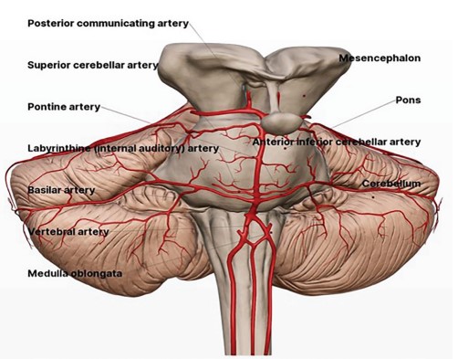
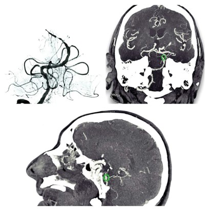
Basilar Artery Syndrome (BAS), also known as Rostral Brainstem Infarction, accounts for 1% of all strokes, with an annual incidence of four patients per 100,000. Top of the Basilar Syndrome (TBS) is characterized by multiple ischemic lesions on diffusion-weighted imaging (DWI) involving more than two territories supplied by branches of the distal basilar artery (cerebellum, hypothalamus, medial diencephalon, and medial thalamus), and occlusion of the distal one-third of the basilar artery (BA) on both time-of-flight magnetic resonance angiography (MRA), and contrast-enhanced MRA. It results from thromboembolic occlusion of the top of the BA. Risk factors include hypertension, diabetes mellitus, obesity, hyperhomocysteinemia, and excessive alcohol intake. Cardiogenic emboli, such as mural thrombi in myocardial infarction, atrial fibrillation, and valvular thrombi in infective endocarditis and valvular heart disease, can also be causative. Hyper coagulable states, like anti phospholipid antibody syndrome, protein C deficiency, and protein S deficiency, may also predispose to thrombotic events. Small vessel vasculitis in sickle cell disease can be associated. Bilateral thalamic ischemia occurs due to occlusion of per for at or vessels. Clinically, patients develop symptoms like visual and oculo-motor defcits and behavioral abnormalities, while motor dysfunction is often absent. This case report describes a 78-year-old man presenting with a generalized seizure and decorticate posturing secondary toTBS, a rare manifestation of a posterior circulation stroke.
References
- 1. Cheng J.H., et al. Characteristics of the ischemic stroke patients whose seizures occur at stroke presentation at a single institution in Eastern China. J Neurol Sci. 2018; 387: 46-50.
- 2. Zhang D., Chen Y., Hao Y., Hu X., He X. Acute basilar artery occlusion presenting with convulsive movements: a systematic review. Front Neurol. 2022 Jan 7; 12: 803618. doi:10.3389/ fneur.2021.803618. PMID: 35069431; PMCID: PMC8777068.
- 3. Otsuji R., Uno J., Motoie R., Karashima S, Ren N., Nagaoka S., Maeda K., Ikai Y., Gi H. Basilarartery occlusion with seizures as a presenting symptom: three cases treated using mechanical thrombectomy. World Neurosurg. 2018 Sep; 117: 32-39. doi:10.1016/j. wneu.2018.05.227. Epub2018 Jun 7. PMID: 29886293.
- 4. Kwak H.S. Park J.S. Mechanical thrombectomy in basilar artery occlusion: clinical outcomes related to posterior circulation collateral score. Stroke. 2020; 51: 2045-2050.
- 5. Algahtani H., et al. Unusual presentation of basilar artery thrombosis. J Cerebrovasc Endovasc Neurosurg. 2020; 22: 282-286.
- 6. Conte W.L., Gill C.E., Biller J. Top of the basilar syndrome presenting with convulsions. JAMA Neurol. 2017; 74(2): 248-249.
- 7. Matsuo K., et al. Top of the basilar syndrome in a young adult initially presenting with a convulsive seizure. Intern Med. 2011; 50: 1425- 1428.
- 8. Wang T.L., et al. Convulsive-like movements as the first symptom of basilar artery occlusive brainstem infarction: A case report. World J Clin Cases 2022; 10(14): 4569-4573.
- 9. Mader E.C. Jr, et al. Stroke-onset seizures during midbrain infarction in a patient with top of the basilar syndrome. J Investig Med High Impact Case Rep. 2020; 8: 2324709620940497.
- 10. Ropper AH. Convulsions in basilar artery occlusion. Neurology. 1988; 38: 1500-1501.
- 11. Bhatt SK, et al. Refractory left focal motor status epilepticus as initial clinical presentation of acute basilar artery thrombosis. Mayo Clin ProcInnovQual Outcomes. 2021: 5: 511-515.
- 12. Inui R., Fujiwara S., Kuroda T., Ohara N., Imamura H., Kohara N., Ariyoshi K., Kawamoto M., Sakai N. Convulsive-like symptoms as initial indications of basilar artery occlusion: a case series study.eNeurologicalSci. 2022 Jun 16; 28: 100410. doi: 10.1016/ j.ensci.2022.100410. PMID: 35757457; PMCID: PMC9218225.
Data Sharing Statement
There are no additional data available.
Funding
This research received no funding.
Author Contributions
All authors contributed significantly to the work and approve its publication.
Acknowledgements
Informed consent was obtained from the patient’s family for publication of this case report. Patient confidentiality and anonymity have been maintained.
Conflicts of Interest
The authors declare no conflicts of interest.
About this article

Cite this article
Pavrey R, Singh R. Basilar artery occlusion and the decorticating patient in the emergency department: a clinical case report. Ind J Emerg Med. 2025;11(2):115-9.
Licence:
Attribution-Non-commercial 4.0 International (CC BY-NC 4.0)This license enables reusers to distribute, remix, adapt, and build upon the material in any medium or format for noncommercial purposes only, and only so long as attribution is given to the creator.
| Received | Accepted | Published |
|---|---|---|
| March 01, 2025 | April 23, 2025 | June 05, 2025 |
DOI: https://doi.org/10.21088/ijem.2395.311X.11225.7
Keywords
Basilar artery syndromeSeizureDecorticate rigidityPosterior circulation stroke.Search for Similar Articles
Similar Articles
- Covid 19 Vaccine: Associated Henoch Schonlein Purpura in Young Adult Female
- A Rare Case of Intolerance to Medically Approved Dose of Sildenafil Citrate in a...
- Acute Mesenteric Ischemia: An Unusual Cause of Hematemesis in a 48 Year Old Man
- Brachial Artery Pseudoaneurysm Related Causally to Iatrogenic Coagulopathy in a...
- Combination Therapy for Acute Management of Yellow Oleander Poisoning: A Case Re...
Article Level Metrics
Last UpdatedSaturday 05 September 2026, 19:26:09 (IST)
4074
Accesses
22
922
00
Citations
NA
NA
NA
Download citation
Article Keywords
Keyword Highlighting
Highlight selected keywords in the article text.
Timeline
| Received | March 01, 2025 |
| Accepted | April 23, 2025 |
| Published | June 05, 2025 |
licence
Attribution-Non-commercial 4.0 International (CC BY-NC 4.0)
This license enables reusers to distribute, remix, adapt, and build upon the material in any medium or format for noncommercial purposes only, and only so long as attribution is given to the creator.