Full Text (PDF)
Indian Journal of Anatomy 14(2):p 69-72, May-Aug 2025. | DOI: https://doi.org/10.21088/ija.2320.0022.14225.5
Case Report
Atypical Foetal Limb Dysplasia: An Anatomical Insight into the Uncommon Lower Limb Congenital Defects
Author Information
Licence:
Attribution-Non-commercial 4.0 International (CC BY-NC 4.0)This license enables reusers to distribute, remix, adapt, and build upon the material in any medium or format for noncommercial purposes only, and only so long as attribution is given to the creator.
Indian Journal of Anatomy 14(2):p 69-72, May-Aug 2025. | DOI: https://doi.org/10.21088/ija.2320.0022.14225.5
How Cite This Article:
Reeha Mahajan. Atypical Foetal Limb Dysplasia: An Anatomical Insight into the Uncommon Lower Limb Congenital Defects. Ind Jr Anat. 2025; 14(2): 69-72.Timeline
Received : July 23, 2025
Accepted : August 06, 2025
Published : August 25, 2025
Abstract
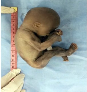
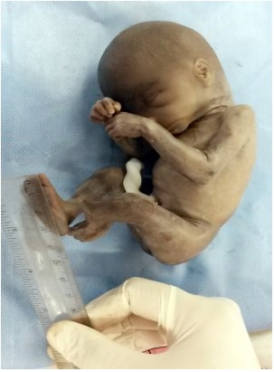
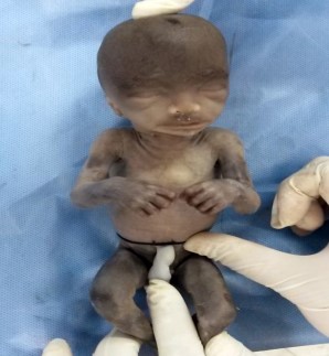
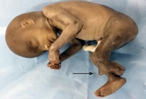
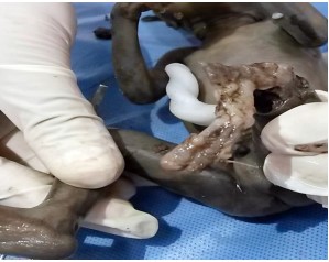
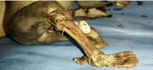
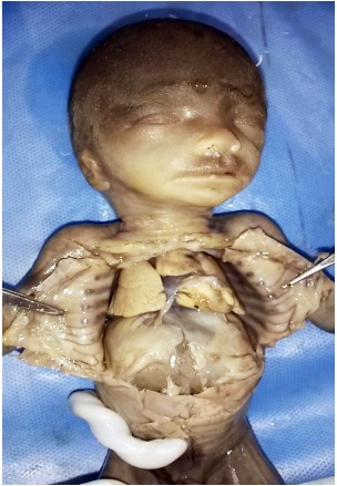
The global birth prevalence of congenital malformations is about 2-3%. Congenital heart defects are the most common followed by the limb anomalies. Congenital limb deficiencies have many causes and often occur as a component of various congenital syndromes. During routine examination of a foetus of 18 weeks gestation obtained for foetal repository in the department of anatomy, some atypical limb anomalies were noted on gross examination which were further evaluated by detailed dissection. This case was identified to be a case of unilateral fibular hemimelia with tibial bone spur and oligodactyly in the same limb. The knowledge of such atypical anomalies is of tremendous clinical relevance for radiologists, obstreticians, paediatricians, paediatric surgeons and orthopaedic surgeons who need to be aware about the presence of such defects at this gestational age for its timely detection, appropriate counselling of parents and timely intervention for surgical planning.
References
- 1. Grogan D.P., Love S.M., Ogden J.A. Congenital malformations of the lower extremities. Orthop Clin North Am. 1987 Oct; 18(4): 537–54.
- 2. Tayel S., Fawzia M., Al-Naqeeb N.A., Gouda S., Al Awadi S., Naguib K. A morpho-etiological description of congenital limb anomalies. Ann Saudi Med. 2005; 25(3): 219–27.
- 3. Bedard T., Lowry R.B., Sibbald B., Kiefer G.N., Metcalfe A. Congenital limb deficiencies in Alberta-a review of 33 years (1980-2012) from the Alberta Congenital Anomalies Surveillance System (ACASS). Am J Med Genet A. 2015 Nov; 167A(11): 2599–609.
- 4. Klaassen Z., Shoja M.M., Tubbs R.S., Loukas M. Supernumerary and absent limbs and digits of the lower limb: a review of the literature. Clin Anat N Y N. 2011 July; 24(5): 570–5.
- 5. Eze K.C., Akhigbe A.O., Awosanya G.O. Fibular Hemimelia: A Case Report. Niger J Clin Pract. 2007; 10(3): 258–61.
- 6. Avila J. Fibular Hemimelia: A Case Series of Nine Patients Treated with Syme Amputation. Glob J Orthop Res [Internet]. 2022 Oct 12 [cited 2025 July 21]; 4(1). Available from: https:// irispublishers.com/gjor/fulltext/fibularhemimelia-a-case-series-of-nine-patientstreated-with-syme-amputation.ID.000577.php
- 7. Baroudi I., Alakhras O., Douri T., Alkhani N. Adams–Oliver syndrome, intestinal obstruction and heart defects: a case series of aplasia cutis congenita. Oxf Med Case Rep. 2022 Jan 24; 2022(1): omab141.
- 8. MSD Manual Professional Edition [Internet]. [cited 2025 July 22]. Congenital Limb Anomalies - Pediatrics. Available from: https://www. msdmanuals.com/professional/pediatrics/ congenital-musculoskeletal-anomalies/ congenital-limb-anomalies
- 9. Yakıştıran B., Altınboğa O., Yüce T., Çağlar AT. Fetal fibular hemimelia with focal femoral deficiency: A case report. Turk J Obstet Gynecol. 2019 Sept; 16(3): 205–7.
- 10. Kavipurapu S.P., Maganthi M., Sundar L.S., Ramya S. Fibular Aplasia, Tibial Campomelia, Oligo-Syndactyly Syndrome and Probable Femur Fibula Ulna Syndrome- Case Reports. J Clin Diagn Res [Internet]. 2021 [cited 2025 July 22]; Available from: https://jcdr.net/article_ fulltext.asp?issn=0973-709x&year=2021&vo lume=15&issue=2&page=SD01&issn=0973- 709x&id=14551
- 11. Bastida M.F., Sheth R., Ros M.A. A BMP-Shh negative-feedback loop restricts Shhexpression during limb development. Development. 2009 Nov 15; 136(22): 3779–89.
- 12. Chiang C. From Oligodactyly to Polydactyly. In: Ruiz i Altaba A., editor. Hedgehog-Gli Signaling in Human Disease [Internet]. Boston, M.A.: Springer US; 2006 [cited 2025 July 23]. p. 137–45. Available from: https://doi. org/10.1007/0-387-33777-6_11
- 13. Cameron T.L., Belluoccio D., Farlie P.G., Brachvogel B., Bateman J.F. Global comparative transcriptome analysis of cartilage formation in vivo. BMC Dev Biol. 2009 Mar 10; 9(1): 20.
Data Sharing Statement
There are no additional data available. All raw data and code are available upon request.
Funding
This research received no funding.
Author Contributions
All authors contributed significantly to the work and approve its publication.
Ethics Declaration
This article does not involve any human or animal subjects, and therefore does not require ethics approval.
Acknowledgements
We would like to express our gratitude to the patients, their families, and all those who have contributed to this study.
Conflicts of Interest
No conflicts of interest in this work.
About this article

Cite this article
Reeha Mahajan. Atypical Foetal Limb Dysplasia: An Anatomical Insight into the Uncommon Lower Limb Congenital Defects. Ind Jr Anat. 2025; 14(2): 69-72.
Licence:
Attribution-Non-commercial 4.0 International (CC BY-NC 4.0)This license enables reusers to distribute, remix, adapt, and build upon the material in any medium or format for noncommercial purposes only, and only so long as attribution is given to the creator.
| Received | Accepted | Published |
|---|---|---|
| July 23, 2025 | August 06, 2025 | August 25, 2025 |
DOI: https://doi.org/10.21088/ija.2320.0022.14225.5
Keywords
Fibular hemimeliaOligodactylyEmbryologyCongenital anomaliesSearch for Similar Articles
Similar Articles
- An Accessory Inferior Pulmonary Vein: A Variation in the Pulmonary Venous Format...
- The Purpose and Utility of the Nipple-Areolar Complex of the Mammary Gland with...
- Morphometric Study of Anterior Border of Human Hip Bone in the Maharashtra Regio...
- Surgeons’ Perspectives on Cadaveric Dissection in Medical Education: A Cross-Sec...
- Transforming Anatomy Education: Emerging Technologies, Challenges, and Future Di...
Article Level Metrics
Last UpdatedTuesday 20 October 2026, 05:48:53 (IST)
6206
Accesses
2
706
00
Citations
NA
NA
NA
Download citation
Article Keywords
Keyword Highlighting
Highlight selected keywords in the article text.
Timeline
| Received | July 23, 2025 |
| Accepted | August 06, 2025 |
| Published | August 25, 2025 |
licence
Attribution-Non-commercial 4.0 International (CC BY-NC 4.0)
This license enables reusers to distribute, remix, adapt, and build upon the material in any medium or format for noncommercial purposes only, and only so long as attribution is given to the creator.