Full Text (PDF)
Physiotherapy and Occupational Therapy Journal 16(1):p 51-54, January-March 2023. | DOI: https://doi.org/10.21088/potj.0974.5777.16123.4
Review Article
Advanced Body Composition Assessment: From Body Mass Index to Body Composition Profiling
Ravi Kumar Chittoria, Bharath Prakash Reddy. J, Jacob Antony Chakiath
Author Information
Licence:
Attribution-Non-commercial 4.0 International (CC BY-NC 4.0)This license enables reusers to distribute, remix, adapt, and build upon the material in any medium or format for noncommercial purposes only, and only so long as attribution is given to the creator.
Physiotherapy and Occupational Therapy Journal 16(1):p 51-54, January-March 2023. | DOI: https://doi.org/10.21088/potj.0974.5777.16123.4
How Cite This Article:
Bharath Prakash Reddy. J, Ravi Kumar Chittoria, Jacob Antony Chakiath/Advanced body Composition Assessment: From body Mass Index to Body Composition Profiling/Physiotherapy and Occupational Therapy Journal. 2023;16(1): 51-54.Timeline
Received : December 17, 2022
Accepted : February 22, 2023
Published : March 25, 2023
Abstract
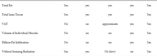
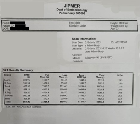
This paper includes a quick overview of common non-invasive techniques in addition to a more in-depth analysis of a body composition assessment method. a technique for figuring out composition using quantitative MRI and using fat as a reference. For whole body measures of adipose tissue (AT), often known as fat, and lean tissue (LT), DXA and quantitative MRIs show excellent agreement, with linear correlations of 0.99 and 0.97 and coefficients of variation (CV) of 4.5 and 4.6% for fat (calculated from AT) and LT, respectively. With a CV of more than 20%, visceral adipose tissue showed much lower agreement. Because of its ability to assess ectopic fat, muscle volumes, and muscle AT infiltration, as well as its speed, quantitative MRI is made possible.
References
- 1. Wang ZM, Pierson RN, Heymsfield SB. The five-level model: a new approach to organizing body-composition research. Am J Clin Nutr 1992; 56:19–28.
- 2. Gallagher D, Heymsfield SB, Heo M, et al. Healthy percentage body fat ranges: an approach for developing guidelines based on body mass index. Am J Clin Nutr 2000; 72:694– 701.
- 3. Thomas EL, Frost G, Taylor-Robinson SD, et al. Excess body fat in obese and normal-weight subjects. Nutr Res Rev 2012; 25:150–61.
- 4. Shen W, Wang Z, Punyanita M, et al. Adipose tissue quantification by imaging methods: a proposed classification. Obes Res 2003; 11:5–16.
- 5. Snyder WS Report of the Task group on reference man: a report Oxford Pergamon 1975;1975.
- 6. Britton KA, Fox CS, Depots EF, et al Circulation 2011;124: e837–e841.
- 7. Demerath EW, Reed D, Rogers N, et al. Visceral adiposity and its anatomical distribution as predictors of the metabolic syndrome and cardiometabolic risk factor levels. Am J Clin Nutr 2008; 88:1263–71.
- 8. Liu J, Fox CS, Hickson DA, et al. Impact of abdominal visceral and subcutaneous adipose tissue on cardiometabolic risk factors: the Jackson Heart Study. J Clin Endocrinol Metab 2010; 95:5419–26.
- 9. Neeland IJ, Ayers CR, Rohatgi AK, et al. Associations of visceral and abdominal subcutaneous adipose tissue with markers of cardiac and metabolic risk in obese adults. Obesity 2013;21: n/a–E447.
- 10. Stahn A, Terblanche E, Gunga H-C. Use of bioelectrical impedance: general principles and overview. In: Preedy VR, ed. Handbook of anthropometry: physical measures of human form in health and disease. New York, NY: Springer New York, 2012:49–90.
- 11. Garg MK, Kharb S. Dual energy X-ray absorptiometry: pitfalls in measurement and interpretation of bone mineral density. Indian J Endocrinol Metab2013; 17:203–10.
- 12. Kramer H, Pickhardt PJ, Kliewer MA, et al. Accuracy of liver fat quantification with advanced CT, MRI, and ultrasound techniques: prospective comparison with MR spectroscopy. AJR Am J Roentgenol 2017; 208:92–100.
- 13. Lareau-Trudel E, Le Troter A, Ghattas B, et al. Muscle quantitative MR imaging and clustering analysis in patients with facioscapulohumeral muscular dystrophy type 1. PLoS One 2015;10: e0132717–16.
- 14. Silver HJ, Niswender KD, Kullberg J, et al. Comparison of gross body fat-water magnetic resonance imaging at 3 Tesla to dual-energy X-ray absorptiometry in obese women. Obesity 2013; 21:765–74.
- 15. Ulbrich EJ, Nanz D, Leinhard OD, et al. Whole-body adipose tissue and lean muscle volumes and their distribution across gender and age: MR-derived normative values in a normal-weight Swiss population. Magn Reson Med2018;79.
Data Sharing Statement
There are no additional data available. All raw data and code are available upon request.
Funding
This research received no funding.
Author Contributions
All authors contributed significantly to the work and approve its publication.
Ethics Declaration
This article does not involve any human or animal subjects, and therefore does not require ethics approval.
Acknowledgements
We would like to express our gratitude to the patients, their families, and all those who have contributed to this study.
Conflicts of Interest
No conflicts of interest in this work.
About this article

Cite this article
Bharath Prakash Reddy. J, Ravi Kumar Chittoria, Jacob Antony Chakiath/Advanced body Composition Assessment: From body Mass Index to Body Composition Profiling/Physiotherapy and Occupational Therapy Journal. 2023;16(1): 51-54.
Licence:
Attribution-Non-commercial 4.0 International (CC BY-NC 4.0)This license enables reusers to distribute, remix, adapt, and build upon the material in any medium or format for noncommercial purposes only, and only so long as attribution is given to the creator.
| Received | Accepted | Published |
|---|---|---|
| December 17, 2022 | February 22, 2023 | March 25, 2023 |
DOI: https://doi.org/10.21088/potj.0974.5777.16123.4
Keywords
BodyMassCompositionSearch for Similar Articles
Similar Articles
- Leadership in Physiotherapy: A Narrative Review
- Effectiveness of Low Level Laser Theapy in the Management of Temporomandibular J...
- The Impact of Air Pollution on Cardiopulmonary Health: Molecular Mechanisms, Pre...
- Efectiveness of Physiotherapy in Postpartum Women with Diastasis Recti: A Review
- Pain Neuroscience Education: Transforming Physiotherapy Practice
Article Level Metrics
Last UpdatedFriday 23 October 2026, 14:01:39 (IST)
4355
Accesses
9
739
00
Citations
NA
NA
NA
Download citation
Article Keywords
Keyword Highlighting
Highlight selected keywords in the article text.
Timeline
| Received | December 17, 2022 |
| Accepted | February 22, 2023 |
| Published | March 25, 2023 |
licence
Attribution-Non-commercial 4.0 International (CC BY-NC 4.0)
This license enables reusers to distribute, remix, adapt, and build upon the material in any medium or format for noncommercial purposes only, and only so long as attribution is given to the creator.